
What Causes Cutaneous Squamous Cell Carcinoma
Cutaneous squamous cell carcinoma is usually driven by UV skin damage over time; know the warning signs, risks, and care options with no referral.





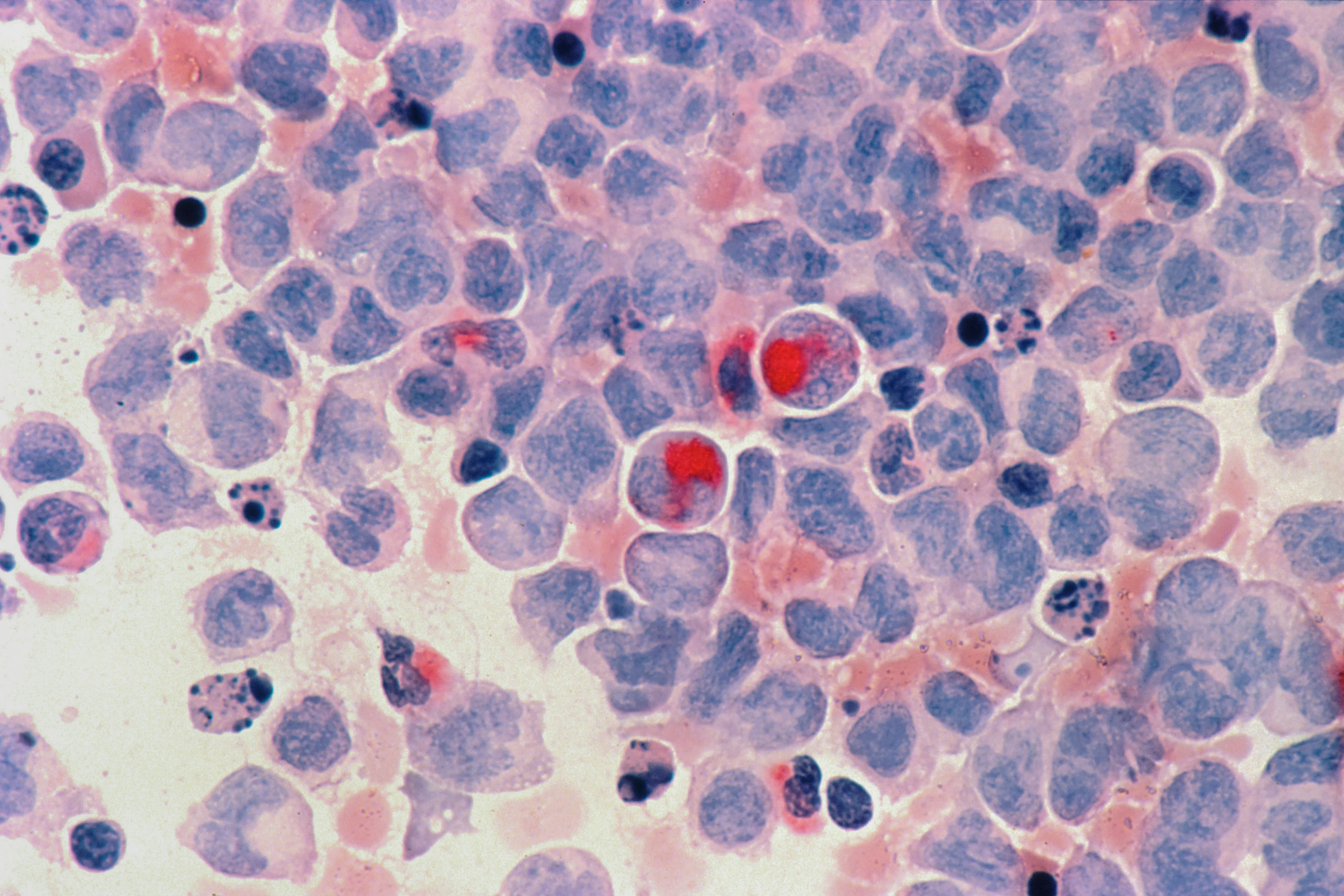
Cutaneous squamous cell carcinoma is a common type of skin cancer that usually starts after years of skin damage, especially from ultraviolet (UV) light. The good news is that when it’s found early, treatment is often straightforward and highly effective. If you’re here because you noticed a new scaly spot, a sore that will not heal, or a crusty bump that keeps coming back, you’re not overreacting. This article walks you through what these spots can look and feel like, what actually causes them in your skin, how diagnosis works (including what a biopsy is like), and what treatment and follow-up typically involve. If you want help deciding how urgent your situation is, PocketMD can help you think it through and plan your next step.
Symptoms and signs to notice
A rough, scaly patch that persists
This often feels like sandpaper and does not smooth out with moisturizer. You might notice it on sun-exposed areas like your face, ears, scalp, forearms, or the backs of your hands. The “so what” is persistence: a spot that keeps returning after it flakes off deserves a closer look.
A sore that will not heal
Some cSCCs look like a small wound that crusts, bleeds, then partly heals, only to open again. That cycle happens because abnormal skin cells keep disrupting normal repair. If a sore has not healed after a few weeks, especially without a clear injury, it is worth getting checked.
A firm bump that crusts or bleeds
You may feel a tender, thickened bump that forms a crust or bleeds with minor friction, like towel drying. It can be skin-colored, pink, red, or darker than your surrounding skin. Bleeding from light contact is a clue that the surface is fragile and abnormal.
A wart-like growth that enlarges
Sometimes it looks like a wart or a “stuck-on” growth, but it keeps growing or changing. The change over time is what matters most, not whether it looks dramatic on day one. If you have something that is clearly bigger month to month, do not wait for it to become painful.
Red flags that need prompt care
Get evaluated quickly if a spot is rapidly enlarging, very painful, or causing numbness or tingling, which can suggest deeper involvement. Swollen, firm lymph nodes near the area—like in your neck, armpit, or groin—also deserve urgent attention. If you are immunosuppressed (for example after an organ transplant), the threshold to be seen should be lower because these cancers can behave more aggressively.
If your clinician wants baseline labs before treatment or to check overall health, Vitals Vault offers a starting from $99 panel with 100+ tests, one visit.
Results in days
Clear next steps
HSA / FSA

Causes and risk factors
Long-term UV exposure damages DNA
The most common driver is ultraviolet light from the sun or tanning beds, which repeatedly injures the DNA inside your skin cells. Over years, your body’s repair systems can miss some of that damage, and abnormal cells can start multiplying. This is why cSCC shows up most often on areas that have seen decades of sun.
Tanning beds raise risk significantly
Indoor tanning delivers concentrated UV that can be intense even if you do not burn. That exposure adds to your lifetime “UV budget,” which is what your skin remembers. If you have used tanning beds in the past, it does not mean you will get cSCC, but it does mean you should be more vigilant about new or changing spots.
Weakened immune system reduces surveillance
Your immune system normally helps spot and remove early abnormal cells, which is a kind of internal quality control. When it is suppressed—after an organ transplant, with certain blood cancers, or from immune-suppressing medications—more abnormal cells can slip through. In that setting, cSCC can grow faster and recur more often, so regular skin checks matter.
Chronic inflammation or scars can trigger it
Skin that is repeatedly irritated or injured can become a site where abnormal cells take hold. Long-standing ulcers, burn scars, or areas of chronic inflammation create an environment of constant repair, and constant repair means more cell turnover. More turnover increases the chance of a “copying mistake” that turns into cancer.
Certain infections and exposures add risk
Some strains of human papillomavirus (HPV) can contribute to squamous cell cancers in specific areas, especially around the genitals or nails. Chemical exposures like arsenic, and prior radiation to the skin, can also increase risk because they damage cells in a different way than UV. The takeaway is not to self-diagnose the cause, but to mention these history details because they change how closely you should be monitored.
How doctors diagnose it
Skin exam with a magnifier
A clinician will look at the spot and the surrounding skin, often using a handheld magnifier with light (dermatoscope). They are looking for patterns that suggest a cancer rather than eczema, a wart, or a benign growth. They will also scan for other sun-damaged areas because cSCC often comes with “neighbors” like precancerous patches.
Biopsy confirms the diagnosis
The only way to know for sure is to sample the spot and look at it under a microscope, which is called a biopsy. Depending on the size and location, this may be a shave, punch, or excisional biopsy, and it is usually done with local numbing medicine. Most people feel pressure more than pain, and the result guides the next step.
Pathology report explains risk level
The pathology report does more than say “yes” or “no.” It can describe how deeply the cancer goes, how aggressive the cells look, and whether there are features linked to higher risk of recurrence. Those details are why two spots that look similar on the surface can get different treatment plans.
Staging and lymph node checks when needed
If the tumor is large, deeply invasive, on high-risk areas like the ear or lip, or you have symptoms like a new lump nearby, your clinician may examine lymph nodes and sometimes order imaging. This is not routine for every small cSCC, and that is reassuring. It is done to make sure treatment is aimed at the full problem, not just what you can see on the skin.
Treatment options
Surgical removal is most common
For many cSCCs, the main treatment is cutting out the cancer with a margin of normal-looking skin. The goal is simple: remove all abnormal cells so they cannot grow back. Your clinician will also think about function and appearance, especially on the face, hands, and lower legs where healing can be trickier.
Mohs surgery for high-risk areas
Mohs surgery removes the cancer in thin layers and checks each layer under a microscope during the visit. It is often used when the spot is on the face, ears, lips, or other areas where preserving healthy tissue matters, or when the borders are hard to see. The “so what” is peace of mind: it offers very high cure rates while sparing as much normal skin as possible.
Scrape-and-burn for selected small lesions
For certain small, superficial tumors, a clinician may scrape the lesion and use heat or electricity to destroy remaining cells, which is called curettage and electrodesiccation. It is quick and done in the office, but it is not the best choice for every location or higher-risk tumor type. If it is offered, ask why it fits your specific spot.
Radiation when surgery is not ideal
Radiation can be used when surgery would be difficult, or as an added treatment if there is concern about leftover cells after removal. It is given over multiple sessions, and the skin can become irritated like a burn during the course. It can be a very practical option for people who cannot tolerate surgery or for tumors in challenging locations.
Systemic therapy for advanced disease
If cSCC has spread or cannot be fully removed, treatments that work through your whole body may be used, including immunotherapy drugs that help your immune system recognize cancer cells. This is less common, but it is important to know it exists because it changes the outlook for advanced cases. If you are in this category, your care team will usually include dermatology and oncology working together.
Living with and after treatment
Expect follow-up skin checks
After you have had one cSCC, your risk of developing another skin cancer is higher because the underlying sun damage is still there. Follow-up visits are not just “busywork”; they are how new spots get caught early, when treatment is simplest. Your clinician will suggest a schedule based on your tumor’s risk features and your immune status.
Do monthly self-skin checks your way
Pick a routine you can actually stick with, like checking your skin after a shower once a month. Use good light and a mirror for your scalp, back, and the backs of your legs, and take a phone photo if you are unsure whether something is changing. The goal is not perfection—it is noticing change sooner than later.
Care for the wound and the scar
Healing depends on location, blood flow, and how much tissue was removed, so your aftercare instructions matter. Keeping the area clean and moist (not dried out) often supports better healing, and protecting it from sun helps the scar fade more smoothly. If you see spreading redness, increasing warmth, pus, or worsening pain, call your clinic because infections are treatable but easier to handle early.
Handle the anxiety of “what if”
It is normal to feel on edge every time you notice a new spot, especially in the first year. Having a plan helps: know what changes trigger a call, and keep your follow-up appointments so you are not carrying the uncertainty alone. If worry is disrupting sleep or daily life, tell your clinician—support is part of cancer care too.
Prevention and lowering your risk
Make sun protection a daily habit
Think of sunscreen as skin maintenance, not a beach-only product. A broad-spectrum SPF 30 or higher, reapplied when you are outdoors, reduces ongoing UV injury that can lead to future cancers. Hats, sunglasses, and tightly woven clothing help because they do not wear off.
Stop tanning beds completely
There is no “safe” tanning bed, because the UV dose is still DNA-damaging even if you do not burn. If you miss the look of a tan, sunless tanning products can give color without UV exposure. This is one of the most powerful risk-reduction steps you can control.
Treat precancerous spots early
Rough sun-damaged patches can be precancers, often called sun spots (actinic keratoses). Treating them does not guarantee you will never get cSCC, but it reduces the number of unstable areas that could progress. If you keep getting these, ask about “field treatments” that treat a whole sun-damaged area rather than one spot at a time.
Extra vigilance if you are immunosuppressed
If you take immune-suppressing medication, your skin needs a tighter safety net. That usually means more frequent professional skin exams and a lower threshold to biopsy anything suspicious. It can also mean coordinating with your prescribing team if you develop repeated or aggressive tumors, because sometimes medication adjustments are possible.
Frequently Asked Questions
What is the main cause of cutaneous squamous cell carcinoma?
Most cases are caused by long-term ultraviolet (UV) damage to your skin, either from the sun or tanning beds. That damage builds up over time, which is why cSCC is more common as you get older. It can also develop in areas of chronic injury or inflammation, especially if your immune system is weakened.
How can you tell squamous cell carcinoma from a harmless scab?
A harmless scab usually has a clear reason (like a cut) and steadily heals over a couple of weeks. A suspicious spot tends to persist, recur, bleed easily, or slowly enlarge even when you leave it alone. If you keep thinking, “Why is this still here?” that is a good reason to get it examined.
Is cutaneous squamous cell carcinoma dangerous or life-threatening?
Many cSCCs are not life-threatening when found early and treated properly, and they can often be cured with a procedure in the office. It becomes more dangerous when it grows deep, involves nerves, recurs, or spreads to lymph nodes. That is why prompt evaluation of changing or non-healing lesions matters.
Does squamous cell carcinoma itch or hurt?
It can, but it does not have to. Some people notice tenderness, burning, or pain when the lesion is bumped, while others only see a rough patch or a sore that bleeds. New pain, rapid growth, or numbness near the spot are reasons to be seen sooner.
Are there blood tests to diagnose cutaneous squamous cell carcinoma?
Diagnosis is usually made with a skin biopsy, not a blood test. Bloodwork may be used as baseline information before certain treatments, or to assess overall health if you have other medical issues. If your clinician recommends labs, Vitals Vault can make it easier to get them done in one visit.