
Preventing Actinic Keratosis What You Need to Know
Preventing actinic keratosis starts with daily UV protection because sun damage triggers these rough precancer spots—get guidance and labs, no referral.





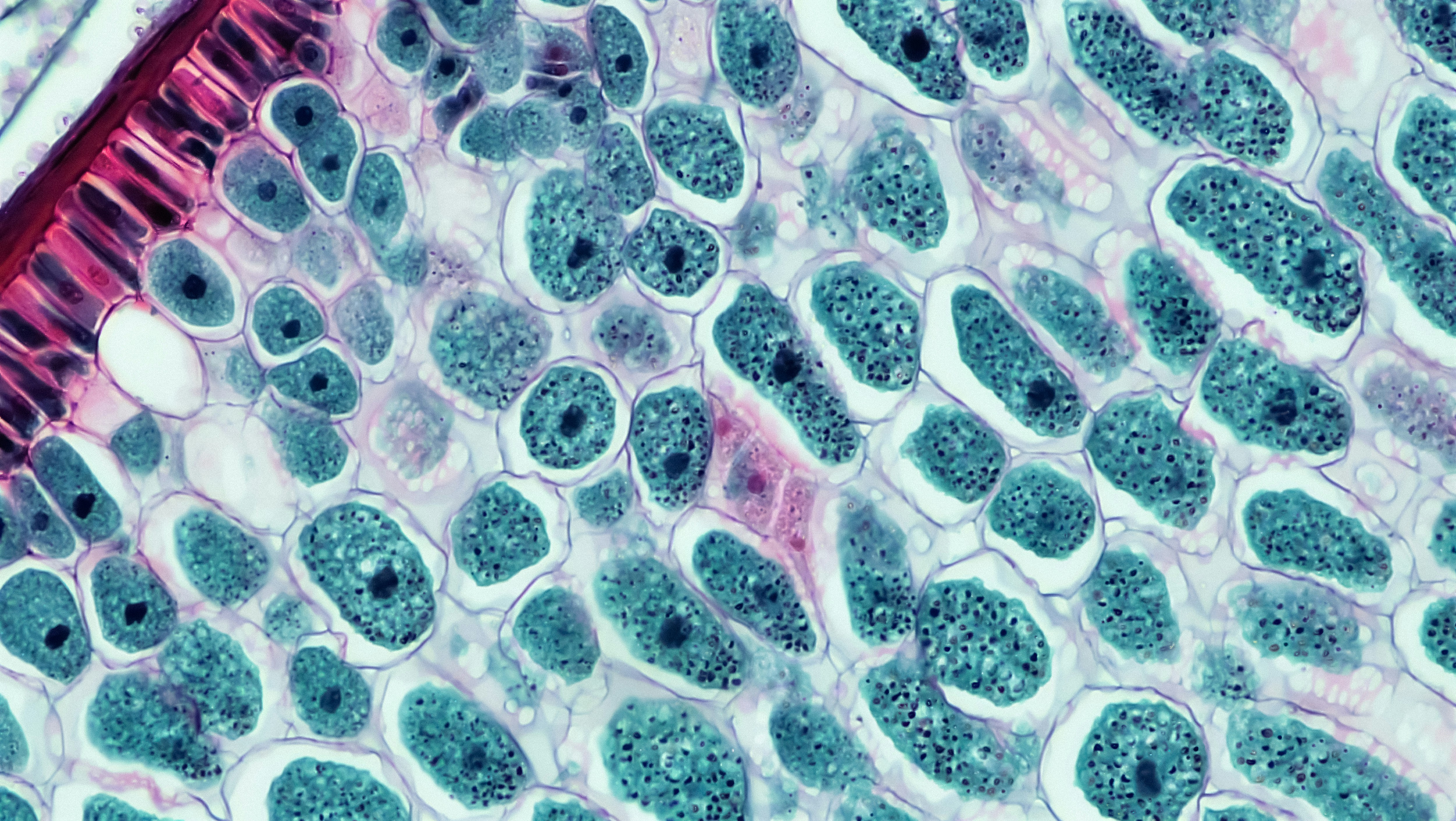
Actinic keratosis is a rough, sun-damaged spot on your skin that can sometimes turn into skin cancer, which is why prevention and early treatment matter. The good news is that your daily UV habits make a real difference, and you can lower your risk starting today. These spots tend to show up on areas that get years of sun, like your face, scalp, ears, forearms, and the backs of your hands. In this guide, you’ll learn what actinic keratosis looks and feels like, who is most at risk, how clinicians confirm the diagnosis, and what treatments and prevention steps are most effective. If you want help sorting out your personal risk or next steps, PocketMD can help you plan what to ask for and when to book a skin check, and Vitals Vault labs can support broader health questions that sometimes come up alongside skin concerns.
Symptoms and signs to watch for
A rough, sandpapery patch
Actinic keratosis often feels rough before it looks obvious, like a tiny patch of sandpaper that won’t smooth out. You might notice it when you wash your face or run your hand over your scalp. That texture matters because it can signal sun-damaged cells that are not behaving normally.
Persistent scaly or crusty spot
Some spots develop visible scale or a thin crust that keeps coming back after you pick at it or moisturize. It may flake, then re-form in the same place. When a spot repeatedly returns, it is worth getting checked rather than assuming it is “dry skin.”
Pink, red, or skin-colored bump
Not every actinic keratosis is brown or dramatic, and many are pink, red, or close to your skin tone. They can look like a small flat patch or a slightly raised bump. This is why “it doesn’t look scary” is not a reliable way to rule it out.
Tenderness, burning, or itching
These lesions can sting, burn, or itch, especially after sun exposure or when you rub them. Discomfort does not automatically mean cancer, but it is your body’s way of telling you the area is irritated and inflamed. If a spot becomes increasingly painful, that is a strong reason to book an exam.
A spot that changes or bleeds
If a sun-exposed spot starts growing quickly, thickening, bleeding with minimal trauma, or forming an ulcer, it needs prompt evaluation. Those changes can overlap with squamous cell skin cancer, which is the main concern with actinic keratosis. If you have bleeding that will not stop, a rapidly enlarging lesion, or a sore that does not heal over a few weeks, do not wait for a routine appointment.
If your clinician is also evaluating fatigue, easy bruising, or immune suppression that could affect healing, Vitals Vault labs can help you start with a starting from $99 panel with 100+ tests, one visit.
Results in days
Clear next steps
HSA / FSA

Causes and risk factors
Years of UV exposure
Actinic keratosis is driven by cumulative ultraviolet (UV) damage, which means it is the total lifetime dose that adds up. Even if you rarely burn now, past sunburns and years of incidental exposure still count. That is why prevention focuses on daily protection, not just “be careful on vacation.”
Fair skin and easy burning
If you have lighter skin, light eyes, or you burn more easily than you tan, you have less natural protection from UV. Your skin cells take damage faster and need more repair, which increases the chance of abnormal patches forming. You can still get actinic keratosis with darker skin, but the risk is generally lower.
Older age and cumulative damage
These spots become more common as you get older because your skin has had more time to accumulate UV injury. It is not “just aging,” though, because the driver is exposure over time. The upside is that changing your habits now still reduces future damage, even if you already have some sun spots.
Outdoor work or high-UV environments
Working outside, living at higher altitude, or spending a lot of time near reflective surfaces like water, sand, or snow increases UV intensity. You may not feel it happening, especially on cool or cloudy days, but UV still reaches your skin. This is why consistent protection during everyday routines is so important.
Weakened immune system
If your immune system is suppressed, your body is less able to clear abnormal skin cells before they become a persistent lesion. This can happen after an organ transplant, with certain medications, or with some chronic illnesses. In that situation, you usually need a lower threshold for dermatology follow-up and more frequent skin checks.
How actinic keratosis is diagnosed
Skin exam in good light
A clinician often diagnoses actinic keratosis by looking and feeling the area, because the rough texture is a key clue. They will also scan nearby sun-exposed skin for other subtle patches you may not have noticed. This matters because treatment decisions change if you have one isolated spot versus a broader “field” of sun damage.
Dermatoscope check for patterns
A dermatologist may use a handheld magnifier with light (dermatoscope) to see patterns beneath the surface. That extra detail helps distinguish actinic keratosis from things like seborrheic keratoses, eczema, or early skin cancers. It is a quick step that can reduce guesswork and unnecessary procedures.
Biopsy when the spot looks atypical
If a lesion is thick, tender, rapidly changing, or bleeding, a small sample may be taken to look at the cells under a microscope (biopsy). This is the most definitive way to rule out squamous cell skin cancer. It can feel intimidating, but it is usually a brief in-office procedure with local numbing.
Your history guides urgency
Your clinician will ask about sun exposure, tanning bed use, past skin cancers, and immune suppression because those details change your risk. They may also ask how long the spot has been there and whether it comes and goes. If you have had prior skin cancer or you are immunosuppressed, it is reasonable to ask how often you should be rechecked.
Treatment options (and what they feel like)
Freezing the spot (cryotherapy)
Freezing is a common in-office treatment for a small number of lesions, and it works by destroying abnormal cells with a short burst of cold. Afterward, the area may blister, scab, and peel over one to two weeks. It is fast, but you may need repeat treatment if the spot returns.
Prescription creams for “field” treatment
When you have multiple rough spots in one area, your clinician may recommend a topical medication that treats the whole sun-damaged zone, not just one lesion. These creams can cause redness, crusting, and soreness for days to weeks, which is uncomfortable but often expected. The payoff is that they can clear visible lesions and also treat early changes you cannot yet see.
Light-activated therapy (photodynamic therapy)
Photodynamic therapy uses a medicine applied to your skin and then a special light to target abnormal cells. The treatment day can involve burning or stinging during the light exposure, and you usually need strict sun avoidance for a period afterward. Many people like it for larger areas because it can treat a broad region with a controlled approach.
Scraping or shaving thicker lesions
If a spot is thicker or has a horn-like scale, your clinician may remove it by gently scraping or shaving it down. This can both treat the lesion and provide tissue for testing when needed. The practical benefit is that it can quickly remove the most suspicious part while you wait for results.
Follow-up to confirm it’s gone
Treatment is not always “one and done,” because sun-damaged skin can produce new lesions over time. A recheck helps confirm the treated area healed as expected and that no suspicious changes are developing. If you keep getting new spots, ask about a prevention-focused plan rather than treating each lesion in isolation.
Living with sun-damaged skin
Build a simple skin-check routine
A monthly self-check helps you notice new rough patches early, when treatment is easier. Pick a consistent day, use bright light, and use a mirror or a partner for your scalp and back. Taking a quick phone photo can help you tell whether a spot is truly changing or just looks different in different lighting.
Protect healing skin after treatment
After freezing or topical therapy, your skin barrier is temporarily weaker, which means it can sting with sun, wind, and harsh products. Gentle cleansing, plain moisturizer, and careful sun protection usually make healing smoother. If you see spreading redness, pus, or increasing pain days after a procedure, call your clinic because infection is uncommon but possible.
Make peace with “maintenance,” not perfection
If you have a lot of cumulative sun damage, the goal is often control and early removal rather than never getting another spot. That mindset shift can reduce anxiety and help you stick with prevention habits. You are not failing if a new lesion appears; you are succeeding if you catch it early.
Coordinate care if you’re high risk
If you are immunosuppressed or you have a history of skin cancer, you may need more frequent dermatology visits and a lower threshold for biopsy. It helps to keep a short list of your medications and past skin treatments so new clinicians can assess your risk quickly. If you are unsure how urgent a new lesion is, PocketMD can help you frame the right questions before you call.
Prevention that actually works
Use sunscreen like a daily medication
Broad-spectrum sunscreen (SPF 30 or higher) works best when it is part of your morning routine, not something you remember only at the beach. Apply it to the places that get incidental sun, like your face, ears, neck, scalp part, and hands. Reapply when you are outdoors for extended periods, because protection fades with time, sweat, and friction.
Cover up with clothing and hats
Clothing is reliable UV protection because it does not wear off the way sunscreen does. A wide-brim hat protects your ears and scalp, which are common sites for actinic keratosis, especially if your hair is thinning. If you work outside, think of sun-protective clothing as your baseline and sunscreen as the backup.
Avoid tanning beds completely
Tanning beds deliver concentrated UV, which accelerates the exact kind of DNA damage that leads to actinic keratosis. Even “just for a base tan” adds risk without offering meaningful protection. If you want the look of a tan, sunless tanners are a safer option because they do not rely on UV injury.
Plan your day around UV peaks
UV is strongest around midday, so shifting outdoor exercise or chores earlier or later can reduce exposure without changing your lifestyle. Shade helps, but it is not perfect because UV reflects off surfaces and reaches you from the sides. Checking the UV index can make the risk feel more concrete, which makes it easier to follow through.
Frequently Asked Questions
Can actinic keratosis go away on its own?
Some spots can fade, especially if you improve sun protection, but many persist or come back in the same area. The bigger issue is that you cannot reliably tell at home which lesions are harmless and which are starting to progress. If a spot keeps returning, thickens, or bleeds, it is worth an exam.
Is actinic keratosis the same thing as skin cancer?
It is not skin cancer, but it is considered a precancer because a small percentage can turn into squamous cell skin cancer over time. That is why clinicians treat it and also focus on preventing new lesions. Think of it as a warning sign that your skin has had enough UV injury to start changing.
What does actinic keratosis look like compared with a normal sunspot?
A typical “sunspot” (freckle-like pigment change) is usually flat and smooth, while actinic keratosis often feels rough or scaly. Color is not a reliable separator because actinic keratosis can be pink, red, tan, or skin-colored. Texture and persistence are often the clues that push clinicians toward treatment or biopsy.
How often should you see a dermatologist if you’ve had actinic keratosis?
The right interval depends on how many lesions you get, your history of skin cancer, and whether your immune system is suppressed. Many people are followed every 6 to 12 months, but higher-risk situations may need closer monitoring. If you notice a new rapidly changing spot between visits, you do not need to wait for your next scheduled check.
Do blood tests help with actinic keratosis prevention?
There is no blood test that diagnoses actinic keratosis, because it is a skin-level problem confirmed by exam and sometimes biopsy. Labs can still be useful if your clinician is evaluating factors that affect healing or immune function, such as medication effects or nutritional issues. If that applies to you, Vitals Vault labs can be a convenient way to start the conversation with results in hand.