
How Is Cutaneous Squamous Cell Carcinoma Diagnosed
Cutaneous squamous cell carcinoma diagnosis starts with a skin exam and biopsy to confirm cancer and guide treatment, with fast care options via PocketMD.





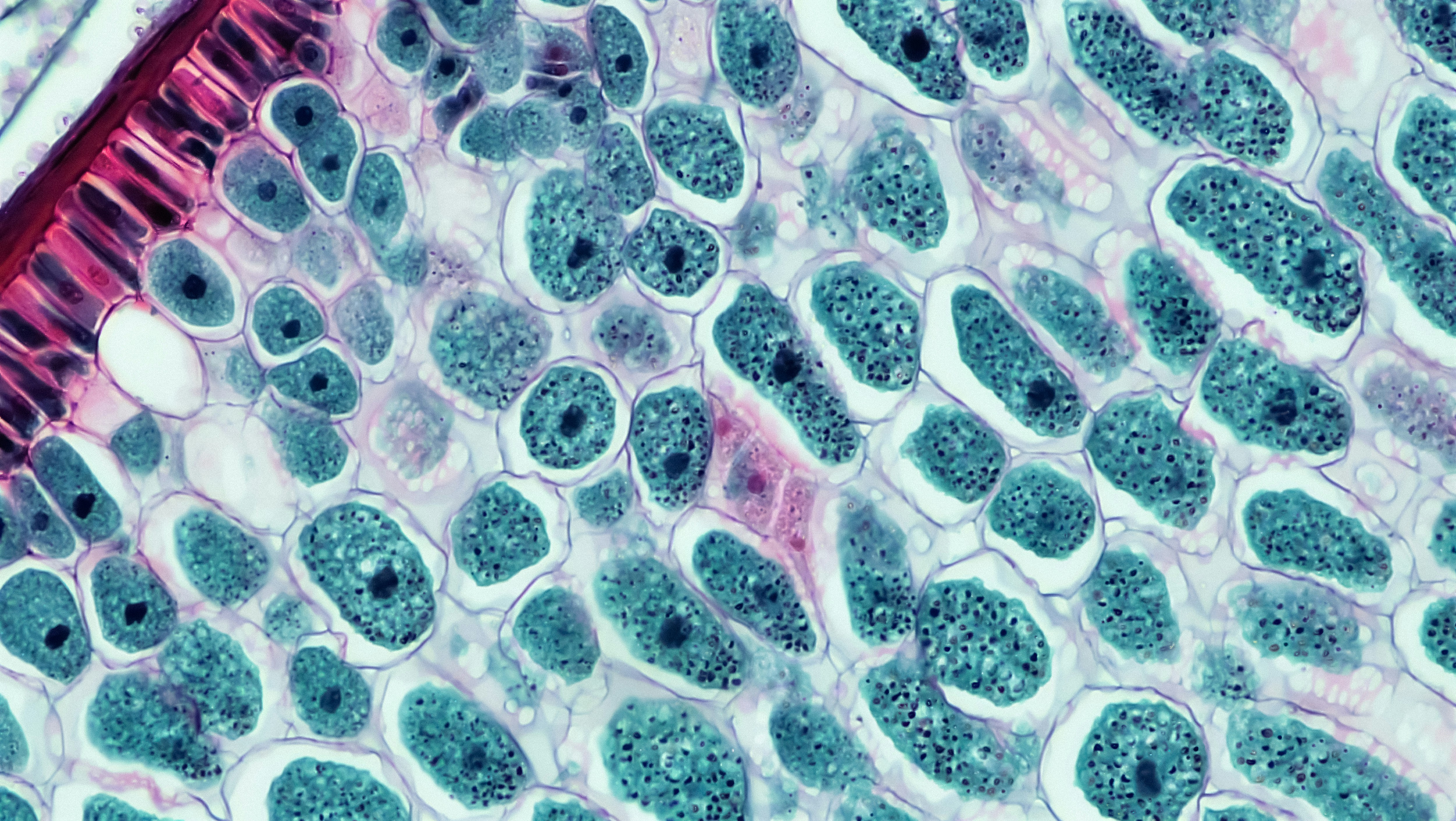
Cutaneous squamous cell carcinoma diagnosis usually comes down to two steps: a careful skin exam and a small skin biopsy that lets a lab confirm what the spot really is. The goal is not just to “name” it, but to figure out how deep it goes, whether it has higher-risk features, and what treatment gives you the best chance of curing it. If you are staring at a sore that will not heal, a scaly patch that keeps coming back, or a fast-growing bump, it is normal to feel anxious while you wait. This guide walks you through what clinicians look for, what a biopsy is like, what pathology results mean, and when imaging or lymph node checks get added. If you want help deciding how urgent your situation is or how to interpret next steps, PocketMD can talk it through with you in plain language.
Symptoms and signs that raise concern
A sore that does not heal
A cut or “scab” that keeps reopening over weeks can be a warning sign because cancers can outgrow the skin’s normal repair process. You might notice it bleeds easily, crusts over, and then comes right back. The “so what” is that persistent non-healing is one of the reasons clinicians move quickly to biopsy instead of trying more creams.
A rough, scaly patch that thickens
Some squamous cell cancers start as a sandpaper-like patch that slowly becomes thicker or more tender. It can look like eczema or a stubborn rash, which is why it gets ignored. If it is growing, changing color, or becoming painful, that change matters more than the exact look.
A firm, growing bump or nodule
A new bump that feels firm and keeps enlarging deserves attention, especially on sun-exposed skin like the face, ears, scalp, and hands. Fast growth can mean the cells are dividing quickly, which sometimes lines up with higher-risk behavior. Even if it does not hurt, the growth pattern is the clue.
Pain, tingling, or numbness nearby
When a skin cancer irritates or grows near small nerves, you can feel burning, tingling, or a deep ache. This can be a sign the tumor is more invasive, which changes how wide the removal needs to be. If you notice new nerve-like symptoms around a lesion, bring it up directly rather than assuming it is “just inflammation.”
Swollen lymph node near the area
A new lump in the neck, armpit, or groin near where the skin spot is located can sometimes mean the cancer has spread to a lymph node. That is not the most common outcome, but it is important because it changes staging and treatment. If you have a rapidly enlarging node, trouble swallowing or breathing, or severe bleeding from a skin lesion, treat it as urgent and get evaluated right away.
If your clinician wants baseline labs before surgery or systemic treatment, you can start with a starting from $99 panel with 100+ tests, one visit.
Results in days
Clear next steps
HSA / FSA

Causes and risk factors
Long-term sun and UV exposure
Most cutaneous squamous cell cancers are driven by DNA damage from ultraviolet light, which builds up over years. That is why they often show up on areas that get “incidental sun” every day, like your ears, lower lip, and backs of your hands. The practical takeaway is that your history of sun exposure matters even if you are careful now.
Tanning beds and intense sunburns
Tanning beds deliver concentrated UV, and repeated exposure can accelerate the kind of DNA damage that leads to skin cancer. A pattern of blistering sunburns also raises risk because it signals high-dose injury to skin cells. If you used tanning beds in the past, tell your dermatologist because it can lower the threshold for biopsy.
Weakened immune system
When your immune system is suppressed, your body is less able to spot and clear abnormal skin cells early. This can happen after an organ transplant, with certain blood cancers, or with immune-suppressing medications. In this setting, squamous cell cancers can behave more aggressively, so clinicians often recommend faster treatment and closer follow-up.
Precancerous spots and chronic irritation
Rough sun-damaged patches called precancers (actinic keratoses) can sometimes progress into squamous cell cancer. Long-standing wounds, scars, or chronically inflamed skin can also be a site where abnormal cells develop. If a “known problem area” changes character—more pain, more bleeding, faster growth—that shift is the reason to re-check it.
HPV and certain high-risk locations
Some squamous cell cancers in genital or peri-anal skin are linked to human papillomavirus (HPV). These areas can be harder to self-check, and symptoms may be subtle at first, like itching or a small sore. The important point is that squamous cell cancer is not only a “sun” disease, so new lesions in covered areas still deserve evaluation.
How doctors diagnose cutaneous squamous cell carcinoma
History and full skin exam
Your clinician will ask how long the spot has been there, how it has changed, and whether it bleeds, hurts, or keeps crusting over. They will also look at the rest of your skin because people often have more than one suspicious lesion at the same time. This visit is where they decide whether to biopsy right away and whether you need a more urgent referral.
Dermatoscopy (magnified skin look)
Many dermatologists use a handheld magnifier with light (dermatoscope) to see patterns under the surface that your eyes cannot pick up. It does not diagnose cancer by itself, but it helps them choose the best spot to sample and avoid missing the most abnormal area. For you, it usually feels like a brief press of a cool lens against your skin.
Skin biopsy to confirm the diagnosis
A biopsy is the definitive step because a pathologist can look at the cells under a microscope and confirm squamous cell carcinoma. Depending on the lesion, you might get a shave biopsy, a punch biopsy, or an excisional biopsy, and all are done with local numbing medicine. The report often includes details like how differentiated the cells are and whether there is nerve involvement, which helps predict risk and guides the treatment plan.
Staging: depth, nerves, nodes, and imaging
If the tumor is large, deep, on a high-risk site (like the ear or lip), or has aggressive features on biopsy, your clinician may check nearby lymph nodes and sometimes order imaging such as ultrasound, CT, MRI, or PET. In select cases, they may recommend a lymph node sampling procedure (sentinel lymph node biopsy) to look for early spread. This is not routine for every small skin cancer, but when it is needed it helps avoid undertreating something that could come back or spread.
Treatment options after diagnosis
Surgical removal with clear margins
For many people, the main treatment is cutting out the cancer with a rim of normal-looking skin around it, which is called getting “clear margins.” The reason margins matter is simple: leftover cancer cells are the most common reason a spot returns. Your clinician will balance taking enough tissue to cure it while protecting function and appearance.
Mohs surgery for high-risk areas
Mohs surgery removes the cancer in thin layers and checks each layer under a microscope during the visit. It is often used on the face, ears, hands, and other areas where you want to spare as much healthy skin as possible while still being confident it is gone. If you are told you need Mohs, it usually means the location or features make precision especially valuable.
Radiation therapy when surgery is hard
Radiation can be used when surgery is not a good fit, such as when a tumor is in a challenging location or when medical issues make surgery risky. It can also be added after surgery if the pathology shows higher-risk features. The tradeoff is that it typically requires multiple sessions and can cause skin irritation and long-term texture changes.
Topical or local treatments for very early disease
When abnormal cells are limited to the top layer of skin (squamous cell carcinoma in situ), your clinician may discuss options like scraping and cautery, freezing, or prescription creams. These approaches can be effective in carefully selected cases, but they are not right for deeper invasive cancers. If your biopsy says “in situ,” ask what makes you a good candidate for a less invasive approach—or why surgery is still recommended.
Systemic therapy for advanced cases
If the cancer has spread or cannot be fully removed, medications that help your immune system recognize cancer cells (immunotherapy) may be used. Some people also need targeted therapy or chemotherapy depending on the situation. Before systemic treatment, clinicians often check baseline blood counts and organ function so dosing is safer, and that is a moment when convenient lab testing can help keep the process moving.
Living with cutaneous squamous cell carcinoma (and follow-up)
Understanding your pathology report
Your report is more than a label, because details like depth of invasion, grade (how abnormal the cells look), and nerve involvement change your follow-up plan. If the report mentions “perineural invasion” (cancer near nerves), that is a reason clinicians may recommend Mohs, wider excision, imaging, or radiation. Bring the report to your visit and ask, “What makes mine low-risk or high-risk?”
Follow-up skin checks that fit your risk
After one squamous cell cancer, your risk of another skin cancer goes up, so follow-up is part of staying well. The schedule depends on your risk factors and the tumor features, but it is often more frequent in the first couple of years. These visits are also when small precancers can be treated early, which can prevent future cancers.
Self-checks without spiraling
A simple monthly scan in good light helps you notice change without checking obsessively. Focus on “new, changing, bleeding, or not healing,” and take a quick photo if something looks different so you are comparing facts, not memory. If you are unsure, it is reasonable to message your clinician with the photo rather than waiting months and worrying daily.
Scar care and recovery after procedures
Most biopsies and surgeries heal well, but the first few weeks can be tender and tight, especially on the face and hands where skin moves a lot. Following wound-care instructions closely lowers infection risk and can improve the final scar. If you notice spreading redness, worsening pain, pus, fever, or a wound that suddenly opens, call your clinician promptly because early treatment prevents bigger problems.
Prevention and lowering your risk going forward
Make sun protection a daily default
Think of sun protection as preventing the next DNA injury, not as a cosmetic habit. A broad-spectrum sunscreen plus hats or UPF clothing is especially important for your ears, scalp part line, and backs of your hands, which are easy to miss. Consistency matters more than perfection, because small daily exposures add up.
Stop tanning beds completely
There is no “safe” tanning bed schedule, because the UV dose is high and the damage is cumulative. If you miss the look of a tan, sunless tanners can give color without UV injury. Quitting now still helps, because it reduces future damage even if you cannot erase past exposure.
Treat precancers early
If you have rough sun spots that your dermatologist calls precancers, treating them is a prevention strategy, not just a cosmetic choice. Early treatment lowers the chance that one of those spots progresses into an invasive cancer. It also gives you a clearer baseline, so new changes stand out sooner.
Extra vigilance if you are immunosuppressed
If you take immune-suppressing medication or have had a transplant, your skin needs a tighter safety net. That often means more frequent professional skin checks and a lower threshold to biopsy anything that changes. It can feel like a lot, but catching problems early is what keeps treatment simpler.
Frequently Asked Questions
Can a doctor diagnose cutaneous squamous cell carcinoma just by looking?
A clinician can often tell when a spot is suspicious, but they cannot confirm squamous cell carcinoma by appearance alone. A biopsy is what makes the diagnosis because it shows the cells under a microscope. The exam still matters because it helps decide how urgent the biopsy is and whether other areas need checking.
What does a skin biopsy feel like, and will it leave a scar?
You usually feel a quick pinch and burn from the numbing medicine, and then pressure but not sharp pain during the biopsy. Most biopsies leave a small scar, although careful wound care can make it less noticeable over time. If the spot is on your face or a high-movement area, ask what scar shape to expect and how to care for it.
How long does it take to get biopsy results back?
Many pathology results come back within several business days, although timing varies by lab and by whether special stains or a second review is needed. Waiting is stressful, so it is reasonable to ask at the time of biopsy when you should expect a call. If you have not heard by that date, follow up rather than assuming “no news is good news.”
If my biopsy says “in situ,” is that still cancer?
“In situ” means the abnormal squamous cells are confined to the top layer of skin and have not invaded deeper tissue. It is often considered an early form of squamous cell cancer, and treating it matters because it can progress over time. The upside is that cure rates are typically excellent when it is addressed promptly.
Do you need blood tests or scans for cutaneous squamous cell carcinoma?
Most small, low-risk skin squamous cell cancers do not require blood tests or scans, because the diagnosis and cure are handled locally with a procedure. Imaging or lymph node evaluation is more likely when the tumor is large, deep, recurrent, on a high-risk site, or shows aggressive features on pathology. If your clinician wants baseline labs before surgery or systemic therapy, a consolidated lab option can help you get it done in one visit.