
How Do Doctors Diagnose Paget Disease of the Breast
Paget disease of the breast is diagnosed with a nipple exam, breast imaging, and a skin biopsy to check for cancer cells—plus labs and no-referral help.





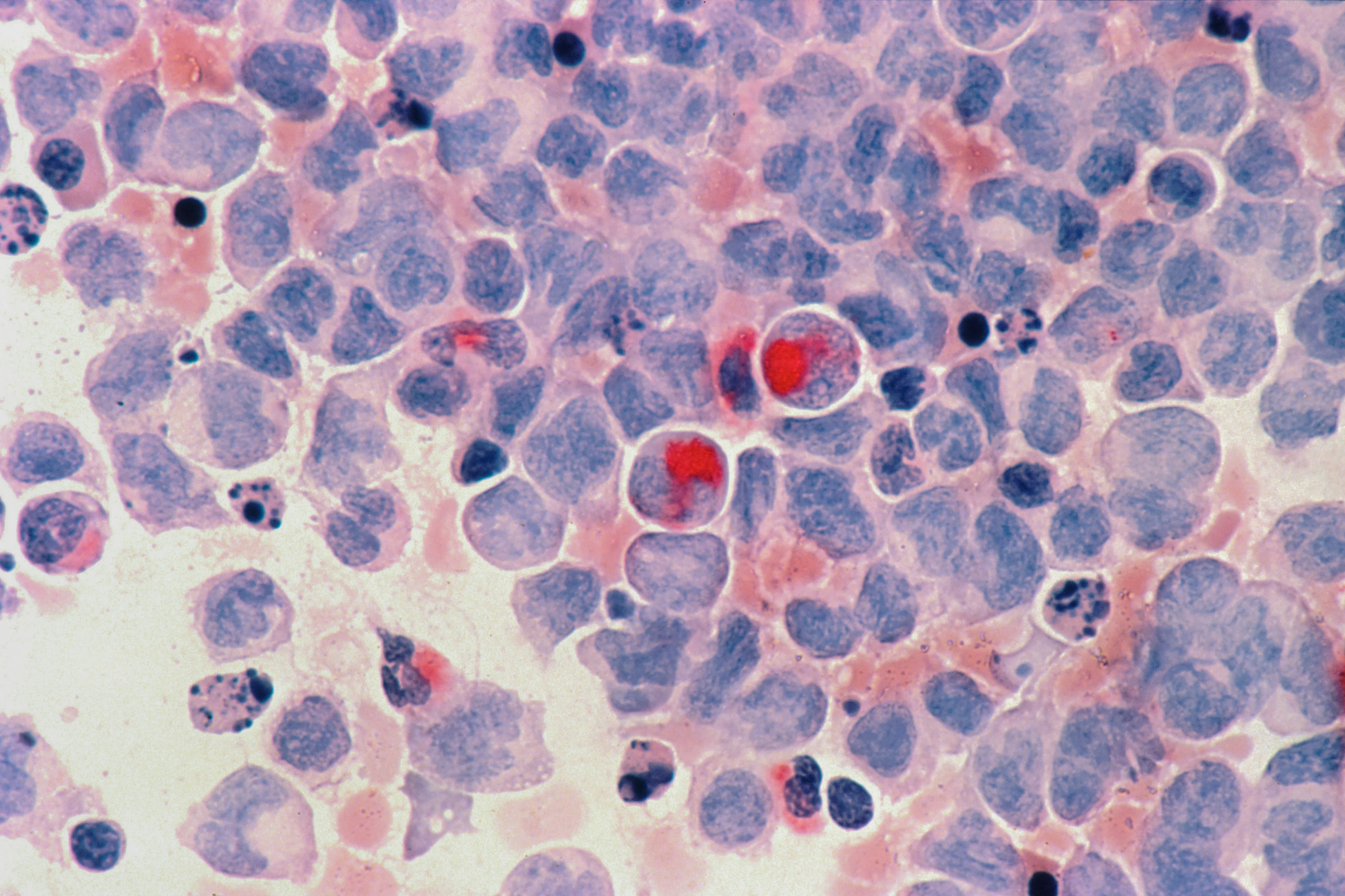
Paget disease of the breast is a rare breast cancer that shows up on the nipple and areola, and doctors diagnose it by looking closely at the skin and confirming it with a small biopsy. The reason this matters is simple: it can look like eczema, but it is often linked to an underlying breast cancer that needs treatment. Most people first notice persistent itching, scaling, or a “raw” nipple that does not fully heal. In this guide, you’ll learn what symptoms make clinicians suspicious, what tests are usually ordered (and why), what results mean, and what treatment decisions typically follow. If you want help making sense of your next step, PocketMD can talk you through the workup, and Vitals Vault labs can support your overall health check-in when you’re coordinating care.
Symptoms and signs that raise suspicion
Persistent nipple rash that won’t clear
If the skin on your nipple or areola stays scaly, flaky, or irritated for weeks despite moisturizers or steroid cream, doctors start thinking beyond simple dermatitis. Paget disease often lingers because the problem is not only on the surface of the skin. The key “so what” is that a rash that keeps coming back deserves a targeted exam and, often, a biopsy.
Itching, burning, or tenderness
You might feel itching that is distracting or a burning sensation that makes clothing uncomfortable. This happens because the nipple skin becomes inflamed and fragile, which can make everyday contact feel sharp. When symptoms are one-sided and persistent, clinicians take it more seriously than typical irritation.
Oozing, crusting, or bleeding
A nipple that weeps fluid, forms crusts, or bleeds easily can be a sign the skin barrier is breaking down. That can happen with infections too, but Paget disease stays stubborn even when you treat it like an infection. If you are soaking through dressings or you see blood, it is a strong reason to be seen promptly.
Nipple flattening or turning inward
If your nipple looks newly flattened or pulled inward, doctors think about changes deeper in the breast that are tugging on the ducts and tissue. This does not prove cancer, but it changes the urgency and the type of imaging you need. It also helps explain why the workup often looks beyond the skin itself.
A breast lump or thickened area
Some people feel a lump, a firm area, or a new difference between breasts. When a lump is present with nipple skin changes, the chance of an underlying breast cancer is higher, so imaging and biopsy decisions move faster. If you also have fever, rapidly spreading redness, or you feel very ill, tell a clinician right away because severe infection needs urgent care too.
If you’re preparing for imaging, biopsy, or surgery, you can check baseline health markers with a starting from $99 panel with 100+ tests, one visit.
Results in days
Clear next steps
HSA / FSA

Causes and risk factors
Cancer cells reaching the nipple skin
In many cases, Paget disease happens when cancer cells from a duct inside your breast travel along the milk ducts to the nipple skin. That is why a skin problem can actually be a clue to something deeper. The practical takeaway is that doctors usually look for an underlying tumor even if you cannot feel a lump.
Underlying duct cancer you can’t feel
Sometimes the associated cancer is “in place” inside the ducts (duct cancer in place [ductal carcinoma in situ]), which can be silent and not form a noticeable mass. You may feel fine except for the nipple changes, which can be confusing and frustrating. Imaging is meant to find these hidden areas so treatment can be properly targeted.
Age and lifetime hormone exposure
Risk rises with age, largely because breast cancer risk rises with age. Longer lifetime exposure to estrogen can also play a role for some people, which is why clinicians ask about your menstrual history and hormone therapy. These questions are not judgment; they help estimate risk and choose the right imaging strategy.
Personal or family history of breast cancer
If you have had breast cancer before, or you have close relatives with breast or ovarian cancer, your clinician may be quicker to order advanced imaging like MRI. The reason is not that symptoms are “worse,” but that the pre-test probability is higher. In some situations, genetic counseling becomes part of the plan because it can affect surgical choices.
Prior chest radiation or high-risk breast lesions
Radiation to the chest earlier in life and certain high-risk biopsy findings can increase your baseline risk of breast cancer. That does not mean Paget disease is inevitable, but it can change how aggressively doctors investigate nipple symptoms. If you know you have a high-risk history, bring it up early so it is not missed.
How doctors diagnose Paget disease of the breast
Focused breast and nipple exam
Your clinician will look closely at the nipple and areola, check both breasts for symmetry, and feel for lumps in the breast and underarm. They will also ask how long the changes have been present and what treatments you have tried, because true Paget disease usually does not fully resolve with typical eczema care. This visit is also where you should mention any nipple discharge, new pain, or a rapidly changing area.
Breast imaging: mammogram and ultrasound
Most people get a mammogram, and many also get an ultrasound, because these tests can spot masses, calcifications, or duct changes that you cannot feel. Imaging helps answer the big question: is there an underlying cancer, and where is it located? Even if imaging looks normal, doctors may still proceed to biopsy because Paget disease can be present without obvious findings.
MRI when imaging is unclear
A breast MRI is often used when the mammogram and ultrasound do not explain your symptoms, or when doctors need a clearer map of how much tissue is involved. MRI is sensitive, which means it can pick up subtle changes, but it can also find “false alarms” that need follow-up. The point is not to overwhelm you with tests; it is to avoid missing a hidden cancer that would change treatment.
Nipple skin biopsy confirms the diagnosis
The definitive step is taking a small sample of the nipple or areola skin (skin sample [biopsy]) and having a pathologist look for Paget cells. This is the test that separates Paget disease from eczema, psoriasis, or infection, which can look similar from the outside. If you have severe swelling, uncontrolled bleeding, or signs of a spreading infection after any procedure, seek urgent care rather than waiting it out.
Treatment options after diagnosis
Surgery to remove affected tissue
Surgery is usually the main treatment because it removes the Paget disease in the nipple and addresses any underlying cancer. Depending on what imaging and biopsy show, that might mean removing the nipple-areola area with a lumpectomy, or it might mean mastectomy. Your surgeon’s goal is clear margins, which means no cancer cells at the edges of what was removed.
Checking lymph nodes when needed
If there is invasive cancer or a higher risk of spread, doctors may sample lymph nodes in your armpit (sentinel node check [sentinel lymph node biopsy]). This helps stage the cancer and guides whether you need additional treatment after surgery. It can also explain why you might have temporary arm soreness or swelling precautions afterward.
Radiation after breast-conserving surgery
If you have a lumpectomy, radiation is commonly recommended to reduce the chance of the cancer coming back in the breast. Radiation is local treatment, which means it targets the breast area rather than your whole body. Many people notice skin irritation and fatigue during treatment, but those effects are usually temporary and manageable with a plan.
Medicine based on tumor type
If an underlying tumor is hormone-sensitive, anti-hormone therapy may be recommended to lower recurrence risk. If the tumor has certain growth signals, targeted therapy may be used, and some people need chemotherapy when the cancer is higher risk. This is why pathology details matter so much: the “type” of cancer changes the best next step.
Supportive care for skin and pain
Even while the cancer is being treated, you still deserve relief from burning, cracking, and irritation. Gentle cleansing, non-stick dressings, and pain control can make daily life more tolerable and protect fragile skin from infection. Ask for a specific skin-care plan because “just keep it clean” is rarely enough when the nipple is actively inflamed.
Living with the workup and recovery
Getting through the waiting period
The hardest part is often the time between noticing symptoms and getting answers. It can help to write down what you are seeing day to day, including photos, because it gives your clinician a clearer timeline. If anxiety is spiking, ask directly when results will be back and who will call you, so you are not stuck refreshing a portal.
Questions to bring to appointments
You will get more clarity if you ask, “What are you trying to rule in or rule out with this test?” and “If it is Paget disease, what is the next step?” You can also ask whether your imaging was able to see behind the nipple well, because that area can be tricky. Bringing a friend or recording the plan (with permission) can help you remember details when you are stressed.
Caring for your nipple skin day to day
Soft bras, breathable fabrics, and non-adhesive dressings can reduce friction, which often makes symptoms flare. Avoid harsh soaps and repeated scrubbing because it can keep the skin in a constant cycle of injury. If you are using any creams, tell your clinician what they are, because some products can mask the appearance of the rash during exams.
Recovery and body image support
After surgery, it is normal to have a mix of relief and grief, even when treatment is going well. Scar care, physical therapy exercises, and reconstructive options can all be part of your recovery plan, but you should not have to guess what is available. If you feel numbness, tightness, or arm swelling, bring it up early because early rehab can prevent long-term limitations.
Prevention and early detection
Don’t ignore a one-sided nipple rash
You cannot fully prevent Paget disease, but you can shorten the time to diagnosis. A rash that is mostly on one nipple and persists beyond a couple of weeks deserves medical evaluation, especially if it is crusting or bleeding. Early attention matters because treatment is simpler when cancer is found at an earlier stage.
Keep up with recommended breast screening
Routine mammograms are designed to find breast cancer before you can feel it, which is relevant because Paget disease is often linked to an underlying tumor. Screening schedules depend on your age and risk, so the “right” interval is personal. If you have a strong family history, ask whether you qualify for earlier or additional imaging.
Know what can mimic Paget disease
Eczema, allergic reactions, and infections can all affect the nipple, and they often improve with appropriate skin treatment. Paget disease tends to persist or recur, which is why follow-up matters if symptoms do not behave like typical dermatitis. The goal is not to panic at every itch, but to notice patterns your body is telling you.
Reduce overall breast cancer risk where you can
Some risk factors are not changeable, but lifestyle choices still matter for your overall breast health. Maintaining a healthy weight, limiting alcohol, and staying physically active can modestly lower breast cancer risk over time. If you are considering hormone therapy, ask about your personal risk profile so the benefits and risks are weighed with your history in mind.
Frequently Asked Questions
Can Paget disease of the breast look like eczema?
Yes, and that is why it is often missed at first. Both can cause redness, scaling, and itching, but Paget disease tends to be persistent and usually affects one nipple. If it does not clearly improve with standard eczema treatment, a biopsy is the step that settles it.
If my mammogram is normal, can I still have Paget disease?
Yes. Some people have Paget disease with no obvious mass or calcifications on mammogram, especially early on. Doctors may add ultrasound or MRI, but the diagnosis ultimately comes from a nipple skin biopsy.
What kind of biopsy is done for suspected Paget disease?
Clinicians usually take a small sample of the nipple or areola skin, often with a punch biopsy or a wedge biopsy depending on the area involved. The tissue is examined under a microscope to look for Paget cells. Your team may also biopsy any suspicious area seen on imaging.
Does Paget disease of the breast always mean you have breast cancer?
Paget disease is considered a form of breast cancer, and it is frequently associated with an underlying duct cancer in the breast. That underlying cancer may be in place (not invasive) or invasive, which is why imaging and sometimes lymph node evaluation are part of the workup. The exact type determines treatment and prognosis.
What should I do while I’m waiting for my biopsy results?
Protect the area from friction with soft fabrics and non-stick dressings, and avoid harsh soaps or repeated scrubbing that can worsen cracking. If you develop rapidly spreading redness, fever, or severe pain, get urgent care because infection can complicate healing. If you want help interpreting the plan and what results might mean, PocketMD can walk you through the usual next steps.