
How Do Doctors Diagnose Actinic Keratosis
Actinic keratosis diagnosis starts with a skin exam for sun-damaged rough spots, sometimes dermoscopy or biopsy to rule out cancer—no referral needed.





Doctors diagnose actinic keratosis by looking closely at a rough, sun-damaged spot on your skin and deciding whether it behaves like a “pre-cancer” change. Most of the time, a careful skin exam is enough, but if a spot looks thicker, tender, fast-changing, or just “not typical,” your clinician may use a magnified light (dermoscopy) or take a small sample (biopsy) to make sure it is not squamous cell skin cancer. Actinic keratosis is common if you have years of sun exposure, especially on your face, scalp, ears, forearms, and hands. The tricky part is that these spots can look subtle, and some early skin cancers can look similar, which is why the diagnosis is really about pattern recognition and risk. In this guide, you will learn what actinic keratosis usually looks and feels like, what raises suspicion, what to expect at a visit, and how treatment choices connect to the way the diagnosis is made. If you want help deciding whether a specific spot needs urgent attention, PocketMD can help you think through your next step and what to ask when you call.
Signs that suggest actinic keratosis
A rough patch you feel first
Actinic keratosis often feels like sandpaper before it looks like much. You might notice a spot that catches on a towel or feels dry and gritty when you run your fingers over it. That texture matters because it reflects surface skin cells that are growing in a disorganized way after sun damage.
Pink, red, or skin-colored scaly spot
The spot can be pink, reddish, tan, or close to your normal skin tone, which is why it is easy to ignore. The scale can come and go, especially if you moisturize or pick at it, but the underlying area tends to persist. If it keeps returning in the same place, that persistence is a clue.
Tenderness or stinging in one area
Some actinic keratoses sting, burn, or feel sore when you press on them, even if they are small. That does not automatically mean cancer, but discomfort is one reason clinicians look more carefully. Pain can also happen when a lesion is thicker or inflamed.
A “horny” bump or thick crust
Sometimes the scale builds into a thicker crust or a little spike of hard keratin, which people describe as a tiny horn. When a spot becomes thick, it is harder to tell by sight alone whether it is still an actinic keratosis or has progressed. This is one of the situations where a biopsy is more likely.
New growth, bleeding, or rapid change
If a spot starts bleeding with minor contact, grows quickly, forms an ulcer, or becomes a firm lump, you should get it checked promptly. Those changes can signal squamous cell skin cancer, which can look like an “angry” actinic keratosis at first. If you also have a rapidly enlarging lesion or a sore that will not heal over a few weeks, do not wait for a routine appointment.
If your clinician is also checking for immune issues or medication effects, Vitals Vault labs can support the workup
starting from $99 panel with 100+ tests, one visit
Results in days
Clear next steps
HSA / FSA

Causes and risk factors
Years of UV exposure and sunburns
Actinic keratosis is basically your skin showing the wear-and-tear of ultraviolet (UV) light. The damage builds over time, so it is common even if you are careful now but had a lot of sun in your teens and twenties. That history helps your clinician interpret what they see on your skin today.
Fair skin and easy burning
If you burn easily or have lighter skin, your skin has less natural protection from UV. That does not mean darker skin cannot get actinic keratosis, but the overall risk is lower. Your clinician will also consider how your skin normally reacts to the sun when deciding how aggressive to be with evaluation.
Older age and cumulative damage
These lesions become more common as you get older because the exposure adds up. You may also have multiple spots in a “field” of sun-damaged skin, not just one isolated lesion. That pattern often shifts treatment toward therapies that treat a whole area, not just a single dot.
Weakened immune system
If your immune system is suppressed—such as after an organ transplant, from certain cancer treatments, or from long-term immune-suppressing medicines—actinic keratoses can appear more often and behave more aggressively. In that setting, clinicians have a lower threshold to biopsy because the risk of skin cancer is higher. It also means follow-up intervals are usually shorter.
Outdoor work and tanning beds
Regular outdoor work exposes you to daily UV, and tanning beds deliver concentrated UV that can accelerate damage. The “dose” matters, so even short sessions add up over years. When your clinician asks about your job and tanning history, it is not small talk—it changes how they judge risk.
How doctors diagnose actinic keratosis
A focused skin exam and history
Diagnosis usually starts with your story and a close look at the spot: how long it has been there, whether it changes, and whether it hurts or bleeds. Your clinician will also scan nearby skin for other rough patches because actinic keratosis often comes in clusters. Expect questions about sun exposure, past skin cancers, and any immune-suppressing medications because those details change the level of concern.
Looking for “field” sun damage
Actinic keratosis is often part of a broader area of sun-damaged skin, which can look mottled, thin, or wrinkled. Seeing that background helps the clinician feel more confident that a rough spot fits the pattern. It also helps them plan treatment, since treating only one lesion may not address the surrounding “at-risk” skin.
Magnified exam with dermoscopy
If the diagnosis is not obvious, your clinician may use a handheld magnifier with light (dermoscopy) to see structures your naked eye cannot. This can help distinguish an actinic keratosis from things like a wart, an inflamed age spot, or an early skin cancer. It is quick and painless, and it often reduces unnecessary biopsies while still catching concerning features.
Biopsy when the spot is atypical
A biopsy means taking a small piece of the lesion to look at under a microscope, which is the most definitive way to rule out squamous cell skin cancer. Doctors are more likely to biopsy if the lesion is thick, firm, tender, rapidly growing, ulcerated, bleeding, or not responding to treatment. If you are anxious about the word “biopsy,” it may help to know that it is usually done with numbing medicine in the office and takes only minutes.
Treatment options after diagnosis
Freezing the spot (cryotherapy)
Freezing with liquid nitrogen is one of the most common treatments for a few isolated lesions. The goal is to destroy the abnormal surface cells so healthier skin can regrow. You can expect redness and a blister or scab for a week or two, and the area may temporarily lighten or darken.
Prescription creams for a wider area
When you have multiple lesions or a whole “field” of sun damage, your clinician may recommend a topical treatment that you apply at home. These creams intentionally cause redness and peeling because they are targeting damaged cells, which can look alarming but is often part of the process. The right option depends on where the lesions are and how much downtime you can tolerate.
Light-based therapy (photodynamic therapy)
Photodynamic therapy uses a medicine applied to your skin and then a special light to activate it, which selectively damages abnormal cells. It can be helpful for larger areas like the face or scalp when you want a more uniform treatment. The main downside is that you can be very light-sensitive for a period afterward, so planning and sun avoidance are important.
Scraping or removing a thicker lesion
If a lesion is thick or has a “horny” buildup, your clinician may remove it by scraping and cautery (curettage and electrodesiccation) or by shaving it off. This both treats the spot and can provide tissue for diagnosis if there is any doubt. It is also a practical option when a lesion keeps returning after simpler treatments.
Follow-up skin checks and monitoring
Treatment is not just about getting rid of today’s spots—it is about catching the next one early. Your clinician may recommend regular skin exams, especially if you have had many actinic keratoses or any skin cancer in the past. Taking a few baseline photos at home can also help you notice change without obsessing over every freckle.
Living with sun-damaged skin
How to watch a spot without spiraling
It is reasonable to keep an eye on a lesion between visits, but you do not need to check it ten times a day. Pick a simple routine, like looking once a month in good light and taking a clear photo with a ruler or coin for scale. What you are watching for is change over time, not perfection in the moment.
What to expect after treatment
Many treatments make the area look worse before it looks better, which can be emotionally harder than people expect. Redness, crusting, and temporary discoloration are common, and they do not automatically mean something went wrong. Ask your clinician what “normal healing” looks like for your specific treatment so you are not guessing.
Skin care that supports healing
Gentle cleansing and plain moisturizers can make healing more comfortable, especially when treatments cause peeling. Avoid picking at scabs because it can delay healing and increase scarring. If you are unsure whether a product is irritating your treated skin, simplify your routine for a week and reintroduce items one at a time.
When to re-contact your clinician
Reach out if a treated spot becomes increasingly painful, develops spreading redness, drains pus, or you get a fever, because those can be signs of infection. You should also check back if a lesion does not improve as expected or returns quickly in the same place. Persistent change is exactly what follow-up is for.
Prevention and lowering your risk
Daily sun protection that you will use
The best sunscreen is the one you will actually apply, so aim for something that feels tolerable on your skin. Broad-spectrum SPF 30 or higher helps reduce new damage, which means fewer new rough spots over time. Reapplying matters most on long outdoor days, not just at the beach.
Cover up during peak UV hours
Clothing is reliable protection because it does not wear off the way sunscreen does. A wide-brim hat and UV-protective shirts are especially helpful for the scalp, ears, and forearms, which are common actinic keratosis zones. If you work outdoors, building shade breaks into your day can make a real difference.
Skip tanning beds completely
Tanning beds are not a “safer” kind of sun, and they deliver UV in a way that accelerates skin aging and cancer risk. If you like the look of a tan, sunless tanners can give color without the DNA damage. This one change can meaningfully reduce your future burden of lesions.
Make skin checks a habit
A quick monthly scan helps you notice new rough areas early, when treatment is simpler. Use a mirror for your back and scalp, or ask a partner to help, because those are easy to miss. If you have a history of many lesions or immune suppression, ask your clinician how often you should have professional full-body exams.
Frequently Asked Questions
Can a doctor diagnose actinic keratosis just by looking?
Often, yes. Many actinic keratoses have a classic rough, scaly look and feel on sun-exposed skin, and an experienced clinician can recognize the pattern during an exam. If the spot is thick, tender, bleeding, or changing quickly, they may recommend dermoscopy or a biopsy to be safe.
When do you need a biopsy for actinic keratosis?
A biopsy is more likely when the lesion looks atypical, feels firm, grows rapidly, ulcerates, or does not respond to standard treatment. The point is to rule out squamous cell skin cancer, which can mimic an actinic keratosis early on. The procedure is usually quick and done with local numbing medicine.
What does an actinic keratosis biopsy show?
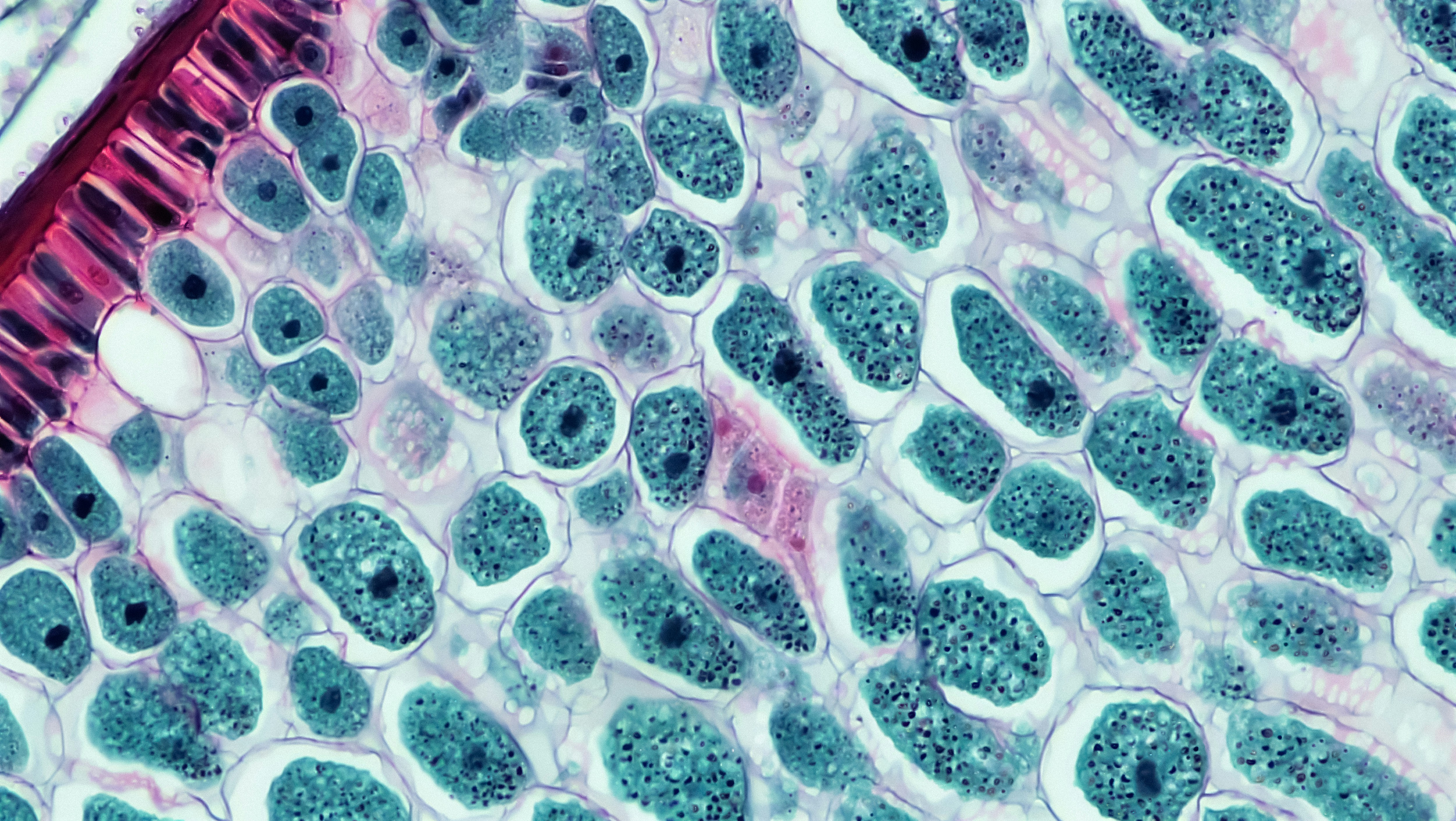
Under the microscope, actinic keratosis shows abnormal growth in the top layers of your skin caused by UV damage. Pathology can also show whether the changes are confined to the surface or have become invasive cancer. That result helps guide whether simple local treatment is enough or whether you need a more definitive removal.
Can actinic keratosis turn into skin cancer?
Yes, some actinic keratoses can progress to squamous cell skin cancer over time, which is why clinicians call them “pre-cancer” lesions. You cannot predict with certainty which individual spot will change, so treatment and monitoring are used to lower risk. If you notice bleeding, fast growth, or a sore that will not heal, get evaluated promptly.
Are there blood tests for actinic keratosis?
No blood test can diagnose actinic keratosis, because it is a skin-level change that is diagnosed by exam and sometimes biopsy. Bloodwork may still be relevant if your clinician is evaluating immune suppression, medication effects, or overall health before certain treatments. If labs are part of your plan, Vitals Vault can make it simpler to get them done in one visit.