
Ductal Carcinoma in Situ Dcis the Breast Cancer You Might Not Have Heard Of
Ductal carcinoma in situ (DCIS) is abnormal cells in a breast duct that haven’t invaded nearby tissue; treatment is tailored, with clear care pathways.





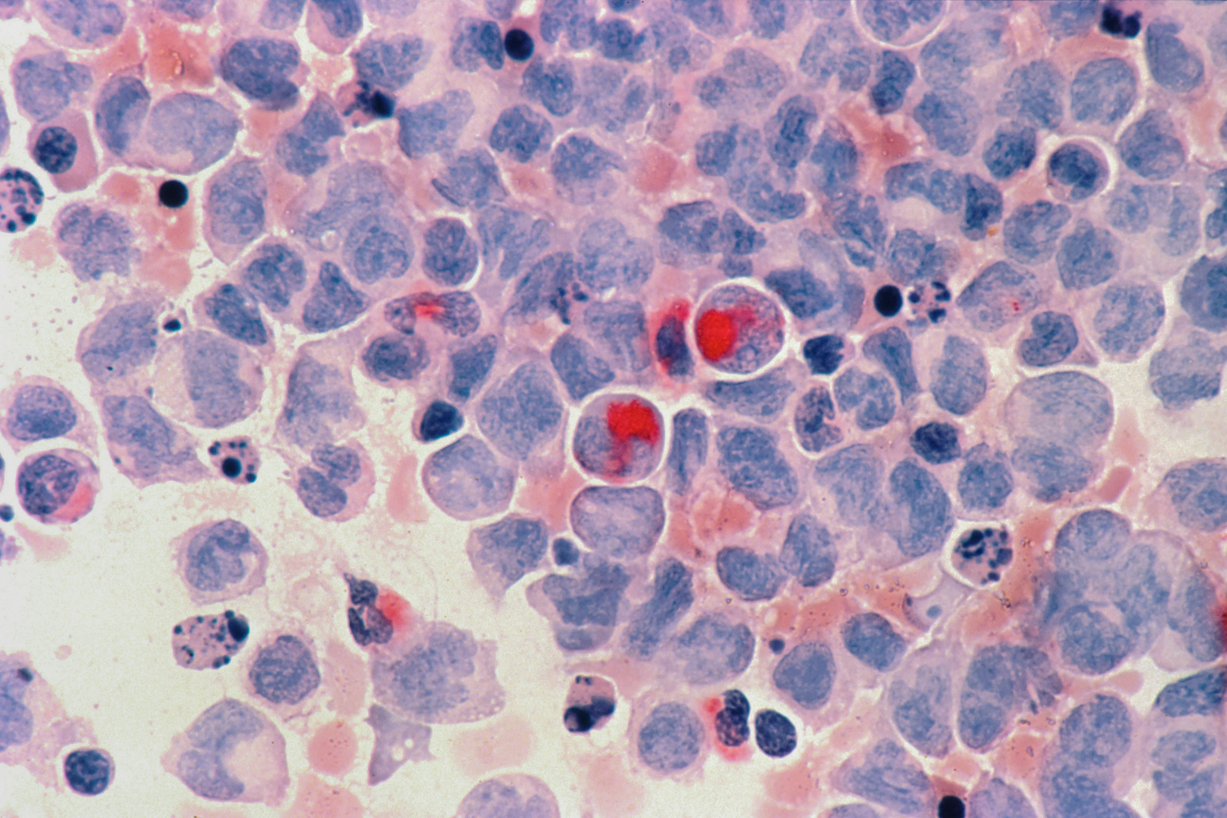
DCIS is a cluster of abnormal cells sitting inside a milk duct in your breast, and the key point is that it has not broken out into nearby breast tissue. That “in place” detail matters because DCIS can often be treated very effectively, but it also creates a confusing gray zone: it is not the same as invasive breast cancer, yet it can become invasive over time. Most people find out they have DCIS after a screening mammogram, not because they felt something wrong. That surprise can be emotionally whiplash—one minute you’re “fine,” and the next you’re hearing the word cancer. This guide walks you through what DCIS is, what (if anything) you might notice, how doctors confirm it, and how treatment choices are usually made so you can ask better questions at your next visit. Because decisions often depend on details from your biopsy report and imaging, it can help to review your results with a clinician who can translate them into plain language. If you want a quick, structured way to talk through options and next steps, PocketMD can help you prepare for those conversations.
Symptoms and signs of DCIS
No symptoms at all
Most of the time, DCIS does not cause a lump or pain, which is why it is commonly found on a screening mammogram. If you feel “normal,” that does not mean the finding is a mistake—it often means the abnormal cells are small and still confined to the duct. The downside is that you may feel blindsided, because your body didn’t give you a warning.
Mammogram shows tiny calcium specks
A classic clue is a pattern of tiny white dots on a mammogram called calcium deposits (microcalcifications). These deposits are not dangerous by themselves, but certain shapes and clusters can signal that cells inside a duct are behaving abnormally. The “so what” is that imaging can raise suspicion, but only a biopsy can confirm DCIS.
A new breast lump or thick area
DCIS usually is not felt, but occasionally you might notice a firm area, especially if there is a larger region involved or there is inflammation around the duct. Any new lump deserves evaluation even if you recently had normal imaging, because not every breast change shows up the same way. If a lump is growing quickly, or the skin looks dimpled or pulled in, you should be seen promptly.
Nipple discharge or crusting
Some people notice discharge, especially if it is spontaneous and coming from one duct, or they notice persistent crusting around the nipple. DCIS can be one cause, but so can benign duct changes, infections, or other growths inside the duct. The important takeaway is to report new, one-sided nipple changes, because they often lead to targeted imaging and sometimes a biopsy.
When to seek urgent care
DCIS itself is not an emergency, but certain symptoms should not wait. If you develop a hot, red, rapidly swelling breast with fever, you could have an infection that needs same-day treatment. If you have chest pain, trouble breathing, or you feel faint, seek urgent care because those symptoms point to problems beyond the breast.
If you’re planning endocrine therapy or want a baseline health check, you can order labs starting from $99 panel with 100+ tests, one visit.
Results in days
Clear next steps
HSA / FSA

Causes and risk factors for DCIS
Random DNA mistakes over time
DCIS starts when cells lining a milk duct pick up DNA changes that make them grow in a more disorganized way. This can happen without a single clear trigger, which is why many people with DCIS have no obvious “cause” to point to. Knowing this can help you stop blaming yourself for something you didn’t control.
Age and lifetime hormone exposure
Your risk rises with age, partly because cells have had more time to accumulate changes. Longer lifetime exposure to estrogen can also play a role, which is why factors like early first period or later menopause can shift risk. This does not mean hormones are “bad,” but it explains why risk is tied to time and biology, not just lifestyle.
Family history and inherited genes
If close relatives have had breast or ovarian cancer, your baseline risk can be higher, and DCIS may be found more often because you are screened more closely. Some inherited gene changes, such as BRCA1 or BRCA2 variants, can raise the chance of developing breast cancers, including lesions that start in ducts. If your diagnosis came at a younger age or you have a strong family pattern, genetic counseling can clarify what it means for you and your relatives.
Dense breast tissue and screening patterns
Dense breast tissue can make mammograms harder to read, but it is also associated with a higher risk of breast cancer overall. People with dense breasts may have more follow-up imaging, which can increase detection of DCIS that might otherwise go unnoticed for longer. The practical point is that your imaging plan may include ultrasound or MRI depending on your density and risk profile.
Prior breast biopsies or chest radiation
A history of certain benign breast changes can be linked to higher future risk, and it often leads to closer surveillance. Radiation to the chest earlier in life, such as for lymphoma treatment, can also increase risk years later because it can damage DNA in breast cells. If either applies to you, your care team may recommend more intensive screening and a lower threshold for biopsy.
How DCIS is diagnosed
Diagnostic mammogram and targeted views
After a screening mammogram raises a question, you usually get a diagnostic mammogram with extra angles and magnified views. This helps the radiologist decide whether the calcifications look suspicious and whether they are in one area or spread out. That map matters because it guides the biopsy approach and can influence surgery planning later.
Breast biopsy confirms what it is
DCIS is diagnosed by looking at tissue under a microscope, usually from a core needle biopsy guided by mammogram (stereotactic biopsy). The biopsy tells you whether the abnormal cells are still confined to the duct and gives details like grade, which describes how aggressive the cells look. If your report mentions “microinvasion” or “invasive,” that changes the conversation, so it is worth asking your clinician to translate every key line.
Pathology details that drive decisions
Your pathology report may include hormone receptor status, meaning whether the cells respond to estrogen or progesterone, because that can open the door to endocrine therapy to lower recurrence risk. It may also mention comedo necrosis, which is a pattern of dead cells in the duct that can be linked with higher-grade disease. These details do not predict your future with certainty, but they help your team weigh how much treatment is likely to help.
Staging, lymph nodes, and red flags
DCIS is considered noninvasive, so lymph nodes are not routinely involved, but sometimes a sentinel lymph node biopsy is done if a mastectomy is planned or if imaging suggests a higher chance of hidden invasion. If you have a rapidly enlarging mass, significant skin changes, or a new hard lump in the armpit, tell your clinician promptly because those findings need faster evaluation. Most people with DCIS will not face those scenarios, but knowing the red flags can keep you from waiting too long.
Treatment options for DCIS
Lumpectomy to remove the area
A lumpectomy removes the DCIS with a rim of normal tissue, aiming for “clear margins,” which means no DCIS at the edge of what was removed. For many people, this is the least body-changing surgical option and preserves most of the breast. The trade-off is that you may need additional surgery if margins are not clear the first time.
Radiation after lumpectomy
Radiation is often recommended after lumpectomy because it lowers the chance that DCIS or an invasive cancer comes back in the same breast. It does not treat your whole body; it is local insurance for the breast tissue that remains. Side effects are usually fatigue and skin irritation, and your radiation oncologist can tell you how your breast size, location of DCIS, and prior radiation history affect the plan.
Mastectomy in selected situations
A mastectomy removes most breast tissue and is considered when DCIS is widespread, appears in multiple areas, or when clear margins are hard to achieve. It can also be chosen for personal reasons, such as wanting the lowest local recurrence risk or avoiding radiation. If reconstruction is on your mind, asking early about timing and options can make the process feel less rushed.
Endocrine therapy for hormone-positive DCIS
If the DCIS is estrogen-receptor positive, medicines that block estrogen’s effect can reduce the risk of DCIS returning and can lower the chance of a new cancer in the other breast. For premenopausal people this is often tamoxifen, while postmenopausal people may be offered an aromatase inhibitor, depending on your situation. The decision is a balance between benefit and side effects like hot flashes, mood changes, or joint aches, so it helps to talk through what you can realistically tolerate.
Active surveillance in research settings
You may hear about “watch and wait” approaches for carefully selected low-risk DCIS, where close monitoring is used instead of immediate surgery. This is still an evolving area and is often offered through clinical trials or specialized programs with strict follow-up. If you are interested, ask what makes a case “low risk,” what imaging schedule would be required, and what would trigger treatment.
Living with DCIS day to day
Handling the emotional whiplash
It is common to feel both relieved and terrified at the same time, because you’re told it is “early” but you still hear the word cancer. Giving your feelings a name—fear of recurrence, fear of overtreatment, fear of missing something—can make decisions clearer. If anxiety is taking over your sleep or appetite, that is a health issue too, and it deserves support.
Questions to bring to your next visit
Ask what grade your DCIS is, whether it is hormone-receptor positive, and what your margin status is after surgery, because those points often shape the next step. It also helps to ask what your estimated risk of recurrence is with and without radiation or endocrine therapy, using your actual pathology. When you hear numbers, ask what they mean in plain language for your life over the next 5 to 10 years.
Follow-up imaging and appointments
After treatment, follow-up usually includes regular mammograms, and the schedule depends on whether you had lumpectomy or mastectomy and whether you still have breast tissue on that side. The goal is not to keep you in a medical loop forever; it is to catch any new changes early, when they are easiest to treat. If you have dense breasts or higher genetic risk, your team may add MRI, and you can ask why it is or isn’t needed for you.
Building a health baseline during treatment
If you start endocrine therapy, tracking symptoms like hot flashes, sleep quality, mood, and joint pain helps you and your clinician decide whether adjustments are needed. Some people also like to check baseline health markers such as lipids and blood sugar, especially if treatment affects activity or weight. If you want that data in one place, VitalsVault lab panels can give a broad snapshot you can review alongside your care plan.
Prevention and risk reduction after DCIS
Stick with your surveillance plan
The most effective “prevention” after DCIS is showing up for the imaging schedule you and your team choose. Recurrences are often found on mammograms before you can feel anything, and early detection keeps treatment simpler. If appointments are hard to manage, ask about coordinating imaging dates months in advance so it becomes routine.
Lifestyle steps that lower overall risk
Regular movement, limiting alcohol, and maintaining a weight that feels sustainable for you are linked with lower breast cancer risk over time. These changes are not a guarantee, but they shift the odds in your favor and often improve energy and sleep during recovery. Pick one change you can keep, because consistency matters more than intensity.
Medication to reduce recurrence risk
If your DCIS is hormone-receptor positive, endocrine therapy can be a risk-reduction tool, not just a “treatment.” The benefit is usually measured as fewer future breast events, but the cost is living with side effects for years. A practical approach is to ask for a clear estimate of benefit in your case and a plan for managing side effects if they show up.
Genetic counseling when risk is high
If you have a strong family history, a diagnosis at a younger age, or relatives with ovarian or pancreatic cancer, genetic counseling can clarify whether an inherited gene change is part of your story. That information can change screening for you and can guide relatives who are wondering what to do. Even when testing is negative, the counseling visit can help you build a risk-based screening plan that feels grounded instead of scary.
Frequently Asked Questions
Is DCIS actually breast cancer or not?
DCIS is abnormal cells inside a milk duct that have not invaded nearby breast tissue, which is why it is sometimes called “stage 0.” People still use the word cancer because the cells can look cancer-like under the microscope and because DCIS can become invasive in some cases. The practical takeaway is that it is highly treatable, but it still deserves a real plan.
Can DCIS spread to lymph nodes or other organs?
By definition, DCIS has not invaded tissue, so it should not spread to lymph nodes or distant organs. The main concern is that a small invasive area can sometimes be found at surgery even if the biopsy showed only DCIS. That is why your team may discuss sentinel lymph node biopsy in certain situations, especially if a mastectomy is planned.
If I have DCIS, will I need chemotherapy?
Chemotherapy is not used for DCIS because there is no invasive cancer to treat throughout the body. Treatment is usually local (surgery, sometimes radiation) and sometimes includes endocrine therapy if the DCIS is hormone-receptor positive. If someone mentions chemo, it usually means invasive cancer was found or there is another diagnosis in the mix.
What are “clear margins,” and why do they matter?
Clear margins mean the edge of the removed tissue does not contain DCIS, which suggests the abnormal area was fully taken out. If margins are close or involved, your risk of the DCIS coming back in that breast is higher, so your surgeon may recommend another surgery to remove more tissue. Asking for the exact margin measurement and what your team considers “adequate” can make the plan feel less mysterious.
What labs should I check if I’m starting tamoxifen or an aromatase inhibitor?
There is no single required blood test that “monitors” these medicines the way some other drugs are monitored, but baseline health labs can still be useful. Many clinicians check things like cholesterol and blood sugar over time, and aromatase inhibitors may prompt attention to bone health through imaging rather than bloodwork. If you want a broad snapshot to share with your clinician, VitalsVault offers panels starting from $99 with 100+ tests in one visit.