
Dealing with Moles When and How to Treat
Dealing with moles means watching for ABCDE changes and symptoms that suggest skin cancer, then choosing safe removal options with no referral.





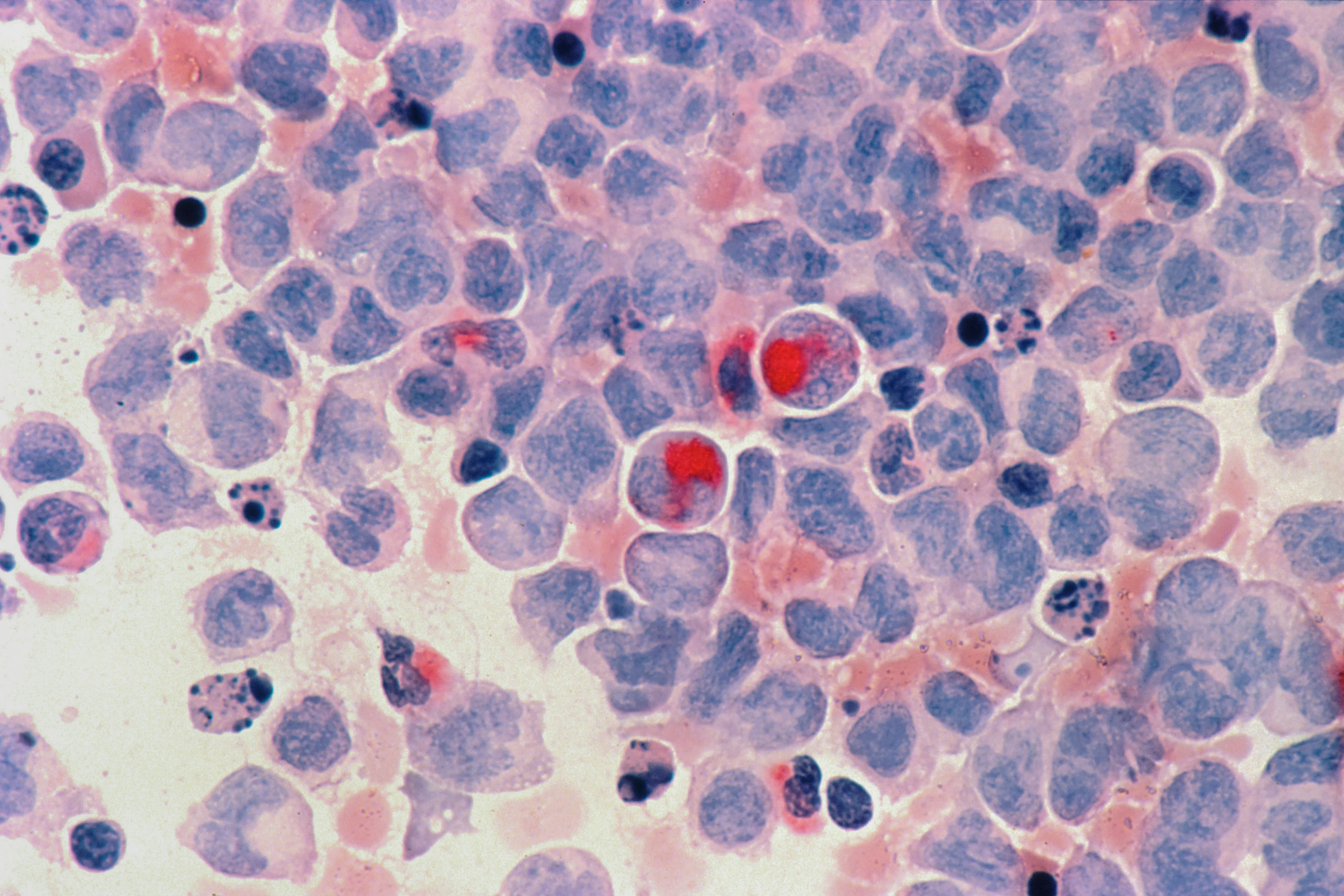
Most moles are harmless clusters of pigment cells, but the reason you pay attention is simple: a changing mole can be an early sign of skin cancer, and catching that early is the whole game. If you are staring at a spot and wondering whether it is “normal,” you are not overreacting. Moles can darken, lighten, or slowly change as you age, during pregnancy, or after sun exposure, and that can be confusing. This guide walks you through what changes matter, when you should get checked, how clinicians decide whether to biopsy, and what removal actually involves. If you want help deciding whether a mole needs urgent attention, PocketMD can talk it through with you in plain language. If your clinician recommends broader health screening, Vitals Vault labs can also support follow-up testing when it fits your situation.
What you might notice with moles
A new mole after age 30
New spots can still be benign later in life, but a brand-new “mole” in adulthood deserves a closer look than one you have had since childhood. What matters is whether it looks different from your other spots and whether it keeps evolving over weeks to months. If it is clearly new and stands out, it is reasonable to schedule a skin exam rather than waiting a year.
ABCDE changes in a mole
The ABCDE rule is a quick way to spot changes that can signal melanoma: uneven shape, irregular border, multiple colors, larger size, or evolution over time. The “E” is the most important because change is often what people notice first. If you see a mole that is actively shifting in shape or color, take a clear photo with a ruler and get it checked.
Itching, pain, or tenderness
Most stable moles do not hurt, so new discomfort can be your body’s way of flagging irritation or inflammation. Sometimes it is as simple as friction from a bra strap, shaving, or a necklace, but persistent tenderness should not be brushed off. If the sensation lasts more than a couple of weeks or keeps returning, a clinician can examine it and decide whether removal is safer than repeated trauma.
Bleeding, crusting, or a sore spot
A mole that bleeds after you nick it while shaving is common, but bleeding without a clear injury is different. Crusting, oozing, or a spot that looks like it is not healing can happen with skin cancers and also with irritated benign growths. If a mole bleeds on its own, forms a scab repeatedly, or looks like an open sore, you should be seen promptly.
The “ugly duckling” mole
You are the best judge of what is normal for your skin, especially if you have many moles. An “ugly duckling” is the one that does not match the rest in color, shape, or texture, even if it does not meet every ABCDE letter. Trust that instinct, because clinicians use the same pattern-recognition approach during skin checks.
If you’re planning a skin check visit, consider baseline labs starting from $99 panel with 100+ tests, one visit to support overall health discussions.
Results in days
Clear next steps
HSA / FSA

Why moles change (and who’s at risk)
Genetics and your baseline mole count
Some people are simply built to make more moles, and that tendency often runs in families. Having many moles is not a diagnosis by itself, but it raises the odds that one could become atypical over time. The practical takeaway is that you benefit from a consistent way to track your skin, such as monthly self-checks and periodic professional exams.
Sun exposure and tanning beds
Ultraviolet light damages the DNA in skin cells, and that damage can show up years later as new moles, changing moles, or skin cancer. A few blistering sunburns, especially in childhood, can meaningfully increase lifetime risk. If you have used tanning beds, it is worth being extra strict about monitoring because the UV dose is intense and concentrated.
Hormone shifts like pregnancy
During pregnancy or other hormone shifts, moles can darken or seem larger, partly because pigment production changes and skin stretches. That can be normal, but it can also hide a real problem if you assume every change is “just hormones.” If a mole is changing quickly, developing multiple colors, or becoming irregular, it still deserves evaluation even if you are pregnant.
Atypical moles and prior skin cancer
If you have had an atypical mole (often called a “dysplastic” mole) removed before, your skin has already shown it can produce spots that look suspicious. A personal history of melanoma or non-melanoma skin cancer raises the stakes further, because recurrence and new cancers are more likely. In that situation, your clinician may recommend more frequent full-body exams and sometimes photo tracking.
Immune suppression and certain medicines
When your immune system is suppressed, your body is less able to spot and clear abnormal cells early. This can happen after an organ transplant, with some autoimmune treatments, or with certain cancers. If you are immunosuppressed, do not wait for a mole to become dramatic before getting it checked, because earlier evaluation is safer.
How clinicians decide what a mole is
Full-skin exam and your history
A good evaluation starts with your story: when you noticed the spot, how it has changed, and whether you have had severe sunburns or a family history of melanoma. Then the clinician looks at your entire skin surface, not just the one mole, because patterns matter. If you can bring a timeline photo, it often makes the decision clearer.
Dermoscopy (a magnified skin look)
Many clinicians use a handheld magnifier with light to see pigment patterns below the surface, which is called dermoscopy. This can help distinguish a harmless pattern from one that looks chaotic or irregular. It is painless, and it often reduces unnecessary biopsies while still catching concerning lesions.
Biopsy: the only way to be sure
If a mole looks suspicious, a biopsy means removing part or all of it so a pathologist can examine the cells under a microscope. That is the definitive step, because you cannot reliably diagnose melanoma by appearance alone. Seek urgent care sooner rather than later if a mole is rapidly enlarging, bleeding without injury, or paired with a new swollen lymph node in the same region.
Mole mapping and photo follow-up
If you have many moles, your clinician may recommend baseline photographs and periodic comparisons, sometimes called total-body photography or mole mapping. This is not about obsessing over every dot; it is about noticing meaningful change early. It can be especially helpful if you have atypical moles, a strong family history, or you struggle to see areas like your back and scalp.
Treatment and removal options (when you actually need them)
Watchful waiting with a tracking plan
If a mole looks clearly benign, the safest “treatment” is often leaving it alone and watching for change. A simple plan helps: take a well-lit photo, include a ruler or coin for scale, and recheck monthly for a few months. This approach keeps you from chasing every normal variation while still catching true evolution.
Shave removal for raised benign moles
For a raised mole that catches on clothing or bothers you cosmetically, a clinician may remove it by shaving it flush with the skin. This can be quick and usually heals with minimal downtime, but it may leave a flat mark and sometimes the mole can regrow if deeper cells remain. If there is any concern for melanoma, clinicians usually avoid a superficial shave and choose a method that captures full depth for accurate diagnosis.
Excisional removal with stitches
When a mole is suspicious or you want the most definitive removal, excision removes the entire lesion with a margin of normal skin and closes the area with stitches. The “so what” is that it gives the pathologist the best sample and lowers the chance of regrowth. It does leave a linear scar, so location and your tendency to form thick scars matter in planning.
If pathology shows melanoma
If the biopsy shows melanoma, treatment depends on how deep it is and whether it has spread. Many early melanomas are treated with a wider excision, and some cases need lymph node evaluation or additional therapies guided by an oncology team. The key point is that early-stage melanoma is often highly treatable, which is why acting on changes is worth the hassle.
Avoid DIY removal and “mole pens”
At-home acids, freezing kits, and so-called mole removal pens can burn the skin and create scars, but the bigger risk is that they destroy the evidence a pathologist needs. If a spot is dangerous, you want a diagnosis, not a cover-up. If a mole is bothering you, a clinician can remove it safely and send it for testing when appropriate.
Living with moles without spiraling
Do a monthly head-to-toe check
A monthly self-check is frequent enough to catch change without turning into daily anxiety. Use a mirror for your back, check your scalp with a comb, and do not forget palms, soles, and under nails. If you find something new, document it and compare in a few weeks rather than relying on memory.
Use photos to reduce guesswork
Your brain is bad at remembering tiny details, especially when you are worried. Taking consistent photos in the same lighting gives you something objective to compare, and it can make a clinic visit more efficient. If you have dozens of moles, consider asking about professional photo tracking so you are not doing it all alone.
Handle irritation and friction smartly
Moles in high-friction areas can get inflamed, which makes them look redder or feel itchy even when they are benign. You can reduce irritation by changing how you shave, using protective bandages for a short period, or adjusting clothing that rubs. If a mole is repeatedly traumatized, removal can be a practical quality-of-life choice.
Know when to book the visit
You do not need an emergency appointment for every freckle, but you also should not wait months for a clearly changing spot. A good rule is to book soon if you see evolution over a few weeks, new bleeding without injury, or a mole that looks unlike your others. If you are unsure, a quick conversation can help you decide whether to monitor or be seen.
How to lower your risk going forward
Make sunscreen a daily habit
Daily broad-spectrum sunscreen helps prevent the UV damage that drives many skin cancers and some mole changes. The habit matters more than perfection, because consistent use adds up over years. If you hate the feel, try a lighter facial formula or a mineral option until you find one you will actually use.
Skip tanning beds completely
Tanning beds are not a “controlled” safer tan; they are a high-dose UV exposure. If you want the look, self-tanner is the safer substitute because it colors the outer skin without DNA damage. This one change can meaningfully lower your long-term risk.
Cover up during peak sun
Clothing is reliable protection because you do not have to remember to reapply it. A wide-brim hat, UV-protective shirts, and seeking shade during midday reduce cumulative exposure, which is what your skin remembers. This is especially helpful if you burn easily or have many moles.
Keep regular skin checks if high-risk
If you have a personal or strong family history of melanoma, many atypical moles, or immune suppression, professional skin exams are part of prevention. The goal is early detection, not finding fault with your skin. Ask your clinician how often makes sense for you, because the right interval depends on your risk profile.
Frequently Asked Questions
How do I know if a mole is cancerous?
You cannot know for sure by looking, but you can spot warning signs like ABCDE changes, bleeding without injury, or a mole that looks unlike your others. A clinician can examine it with magnification and decide if a biopsy is needed. The biopsy is what confirms whether cancer cells are present.
Is it normal for moles to change over time?
Some slow change can be normal as you age, and pigment can shift with sun exposure or hormonal changes. What is not “normal” is rapid evolution, multiple new colors, irregular borders that are getting worse, or spontaneous bleeding. When in doubt, take a photo and schedule a skin check.
Should I remove a mole that gets irritated when I shave?
If a mole is repeatedly nicked or rubbed, it can stay inflamed and look more concerning than it really is. Removal can be a practical option, especially if it keeps bleeding from minor trauma. The safest route is clinician removal so it can be evaluated and, when appropriate, sent to pathology.
Can I remove a mole at home with a mole pen or acid?
It is not a good idea because you can burn your skin, cause infection, and leave a scar that is harder to treat later. More importantly, you can destroy a spot that should have been biopsied, which delays diagnosis. If a mole bothers you, medical removal is quick and gives you clarity.
Do blood tests help diagnose melanoma or suspicious moles?
For a suspicious mole, the key test is a skin biopsy, not a blood test. Blood work can still be useful for your overall health or to prepare for procedures, and your clinician may order labs based on your broader situation. If you want convenient baseline testing, Vitals Vault offers panels starting from $99 with 100+ tests in one visit.