
Are You at Risk for Melanoma 5 Surprising Factors to Consider
Melanoma risk rises when pigment cells mutate, and sun isn’t the only driver. Know 5 surprising risks, warning signs, and next steps—no referral.





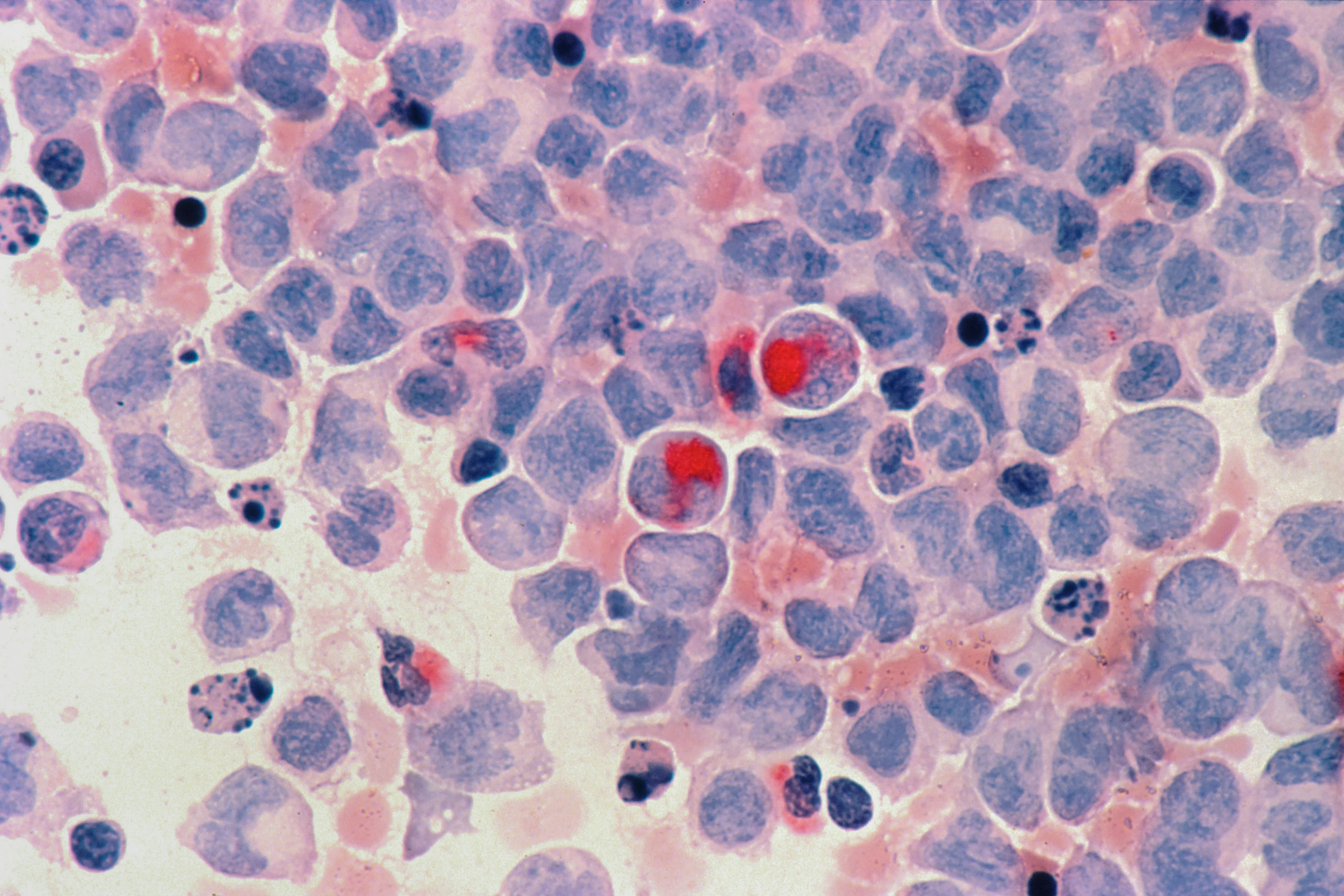
Melanoma is a serious skin cancer that starts in your pigment-making cells, and your risk is shaped by more than just sunburns. The goal is simple: notice suspicious changes early and get the right kind of exam fast, because early melanoma is often highly treatable. In this guide, you’ll learn the warning signs that matter, plus five risk factors people don’t always expect—like certain medications, lots of moles, and immune suppression. You’ll also see what a real diagnosis looks like (hint: it’s not a blood test), what treatment usually involves, and how to protect your skin without living in fear. If you want help deciding how urgent a spot is, PocketMD can help you think through next steps and what to ask for at your visit.
Melanoma signs you can spot
A mole that is changing
The most important clue is change over time, especially if a spot looks different from your other moles. You might notice it getting larger, darker, or more raised over weeks to months. That “new behavior” matters because melanoma is defined by cells that keep growing when they shouldn’t.
The “ugly duckling” spot
Sometimes the red flag is not a specific rule, but the fact that one spot just doesn’t match the rest of your skin. If most of your moles are small and evenly brown, a single larger or more irregular one deserves attention. Trust that instinct, because your eyes are good at noticing patterns even when you can’t explain why.
Irregular border or uneven color
A melanoma can have edges that look jagged or blurred, and it may contain multiple shades rather than one uniform color. You might see brown mixed with black, red, pink, or even bluish tones. This matters because uneven growth in different areas of the spot can show up as uneven pigment on the surface.
Bleeding, crusting, or a sore spot
A mole that bleeds easily, forms a crust, or feels tender is not automatically melanoma, but it should not be ignored. Irritation can happen from shaving or friction, yet melanoma can also become fragile and inflamed. If a spot keeps reopening or won’t heal over a few weeks, it’s worth getting checked.
New dark streak under a nail
A new brown or black band under a fingernail or toenail can be harmless pigment, but it can also be a type of melanoma that starts in nail tissue. It becomes more concerning if the streak is widening, uneven, or pigment seems to extend onto the surrounding skin. If you did not clearly injure the nail and the color persists, don’t wait it out.
If you’re also tracking overall health risks, you can add baseline labs starting from $99 panel with 100+ tests, one visit
separate from skin biopsy decisions
Results in days
Clear next steps
HSA / FSA

Causes and risk factors (including surprising ones)
Indoor tanning counts as high-intensity UV
Tanning beds deliver concentrated ultraviolet (UV) radiation, which can damage the DNA inside your pigment cells. Even if you “don’t burn,” that damage can still accumulate. The so-what is that indoor tanning can raise melanoma risk in a way that is preventable, starting today.
Lots of moles or atypical moles
If you have many moles, you simply have more pigment-cell “real estate” where a problem could start. Atypical moles (dysplastic nevi) can also look irregular even when they are benign, which makes it easier to miss a dangerous change. The practical takeaway is that you benefit from a baseline full-skin exam and photos so future changes are easier to spot.
Family history and inherited risk
If a close relative has had melanoma, your risk can be higher because of shared genes and shared sun-exposure patterns. This matters because you may need earlier or more frequent professional skin checks than someone without that history. It also means you should be extra strict about sun protection for kids in the family, since early-life UV damage can echo later.
Immune suppression (transplants, some meds)
Your immune system helps patrol for abnormal cells, including early cancer cells, and immune suppression can weaken that surveillance. This can happen after an organ transplant, with certain autoimmune treatments, or with long-term immune-suppressing medications. The so-what is that you should treat new or changing skin spots as higher priority, even if you are younger than the “typical” melanoma age.
Photosensitizing meds and “incidental” sun
Some medications make your skin more sensitive to sunlight, which means you can burn faster during everyday exposure like driving, walking the dog, or sitting near a window. A burn is a sign of DNA injury, and repeated injury raises the odds of mutations over time. If you recently started a new medication and notice you are burning more easily, ask your clinician or pharmacist whether sun sensitivity is a known effect and adjust protection accordingly.
How melanoma is diagnosed
A focused skin exam and your history
A clinician starts by looking closely at the spot that worries you and then scanning the rest of your skin for patterns. You’ll be asked about how long it has been there, whether it is changing, and your personal risk factors like tanning bed use or immune suppression. This matters because the story of the spot often determines how quickly you need a biopsy.
Dermatoscope exam (magnified skin view)
A dermatologist may use a handheld magnifier with light (dermatoscope) to see structures under the surface that your naked eye can’t. This can help separate a harmless mole from one that has suspicious pigment networks or irregular growth. It does not “rule out” melanoma by itself, but it helps decide whether a biopsy is the safest next step.
Skin biopsy is the deciding test
Melanoma is diagnosed by removing part or all of the spot and having it examined under a microscope (biopsy). If you are worried about scarring, it helps to know that the bigger risk is delaying a diagnosis when a lesion is truly dangerous. Seek urgent evaluation if a spot is rapidly enlarging, bleeding repeatedly, or paired with new swollen lymph nodes, because those changes should not wait months.
Staging tests if melanoma is confirmed
If melanoma is found, the next question is how deep it is and whether it has spread, which guides treatment. You may hear about depth measurements and sometimes a lymph node procedure (sentinel lymph node biopsy) to check early spread. Imaging is not needed for every case, but it can be used when the melanoma is thicker or symptoms suggest it has traveled.
Treatment options if it is melanoma
Surgical removal with a safety margin
For many early melanomas, the main treatment is cutting out the melanoma plus a rim of normal-looking skin around it to reduce the chance of cells being left behind. This is usually done as an outpatient procedure. The so-what is that early detection often means surgery can be curative without needing more intensive therapy.
Lymph node evaluation when risk is higher
If the melanoma is deeper or has other high-risk features, your team may recommend checking the nearest “first” lymph node (sentinel node) for spread. This helps stage the cancer and can change what treatments are recommended next. Even when the node is negative, the information can give you a clearer follow-up plan.
Immunotherapy for advanced disease
Some melanomas respond to medicines that help your immune system recognize and attack cancer cells (immunotherapy). These treatments can be life-extending and sometimes long-lasting, but they can also trigger immune side effects that feel like inflammation in normal organs. If you are on immunotherapy, new diarrhea, shortness of breath, or severe fatigue should be reported quickly rather than “pushed through.”
Targeted therapy for specific mutations
Some melanomas have specific genetic changes in the tumor that can be targeted with medication, which is why tumor testing may be recommended. When it works, targeted therapy can shrink tumors quickly, although resistance can develop over time. The practical takeaway is that “melanoma” is not one single disease, and treatment is increasingly personalized to the tumor’s biology.
Follow-up plan and skin surveillance
After treatment, you’ll usually have scheduled skin exams and sometimes imaging based on your stage and risk. This is not busywork; it is how recurrences and new melanomas are caught early, when options are best. You can make these visits more effective by bringing photos of any changing spots and a short timeline of when you noticed them.
Living with higher melanoma risk
Do monthly skin checks that you can sustain
A monthly check works because it makes “change” easier to see, but it only helps if you actually do it. Use the same routine each time, including your scalp, soles, and between toes, and use a mirror or a partner for your back. Take a few clear photos of moles you’re watching so you’re not relying on memory.
Know what to do when you find a change
If you spot a change, your next step is usually a timely dermatology visit, not a week of internet spiraling. When you call, say that the lesion is new or changing and mention any high-risk factors like immune suppression or a prior melanoma. If appointments are backed up, ask about a cancellation list or an urgent spot-check visit.
Protect your mental bandwidth
Being vigilant can slide into constant checking, especially if you have anxiety or a family history. It helps to set a schedule and stick to it, because more checking does not always equal more safety. If worry is taking over your day, bring it up at your visit; a clinician can help you calibrate what is “watchful” versus “stuck.”
Coordinate care if you take immune-altering meds
If you are on medications that suppress your immune system, your skin is only one part of the picture. Let your prescribing clinician know about any suspicious lesions and any history of skin cancer, because it can affect medication choices and monitoring. Keeping your medication list updated also helps your dermatologist interpret rashes and pigment changes that are not melanoma.
Prevention that actually lowers risk
Make sunscreen a daily habit
Sunscreen is most effective when it is routine, not just a beach-day product. Use a broad-spectrum option and apply enough to cover exposed skin, then reapply when you are outside for extended periods. The so-what is that preventing burns and chronic UV damage reduces the mutation load your pigment cells accumulate.
Use clothing and shade strategically
Clothing, hats, and shade work even when you forget to reapply sunscreen, which makes them powerful. A wide-brim hat protects areas where melanoma can be missed, like the ears and scalp line. If you plan outdoor time, building shade into the plan is an easy win that does not depend on perfect technique.
Skip tanning beds completely
There is no “safe” tanning bed schedule because the exposure is still UV injury. If you want the look of a tan, sunless tanners can color the outer skin layer without the DNA damage. This swap is one of the most direct ways to lower risk without giving up how you want to look.
Get the right screening for your risk
If you have many moles, atypical moles, a strong family history, or immune suppression, a periodic full-body skin exam can catch problems earlier than self-checks alone. Some people also benefit from total-body photography to track change over time. Screening is not one-size-fits-all, so it’s worth asking what interval makes sense for you.
Frequently Asked Questions
What are the ABCDE warning signs for melanoma?
ABCDE is a quick way to remember common red flags: asymmetry, border irregularity, color variation, diameter that is growing, and evolving over time. The most useful letter is often E, because change is what separates “my normal mole” from “something new.” If a spot is evolving, it deserves a professional look even if it doesn’t hit every letter.
Can you get melanoma on skin that doesn’t see much sun?
Yes. Melanoma can show up on areas that get little sun exposure, including the soles of your feet, under nails, and the genital area. Sun is a major driver, but it is not the only one, which is why full-body checks matter when you are higher risk.
Does melanoma itch or hurt?
It can, but many melanomas do not cause any sensation at all. Itching, tenderness, or bleeding can happen when a lesion is irritated, inflamed, or fragile, and that is why symptoms should raise your attention rather than reassure you. The safest rule is to prioritize change, not comfort level.
Is there a blood test that can detect melanoma early?
A skin biopsy is the test that diagnoses melanoma, because it directly examines the cells in the lesion. Blood tests may be used in some situations after diagnosis to monitor overall health or treatment effects, but they do not replace a biopsy for an early suspicious mole. If you want baseline health labs for other reasons, Vitals Vault lab panels can help, but they are separate from melanoma detection.
How fast should you see a dermatologist for a changing mole?
If a spot is clearly changing over weeks to months, it is reasonable to seek an appointment as soon as you can rather than waiting for your next routine visit. If it is rapidly enlarging, repeatedly bleeding, or you notice new swollen lymph nodes, treat that as urgent and ask for a prompt evaluation. When you call, saying “new or changing lesion” can help the office triage you appropriately.